Genome-edited Cells
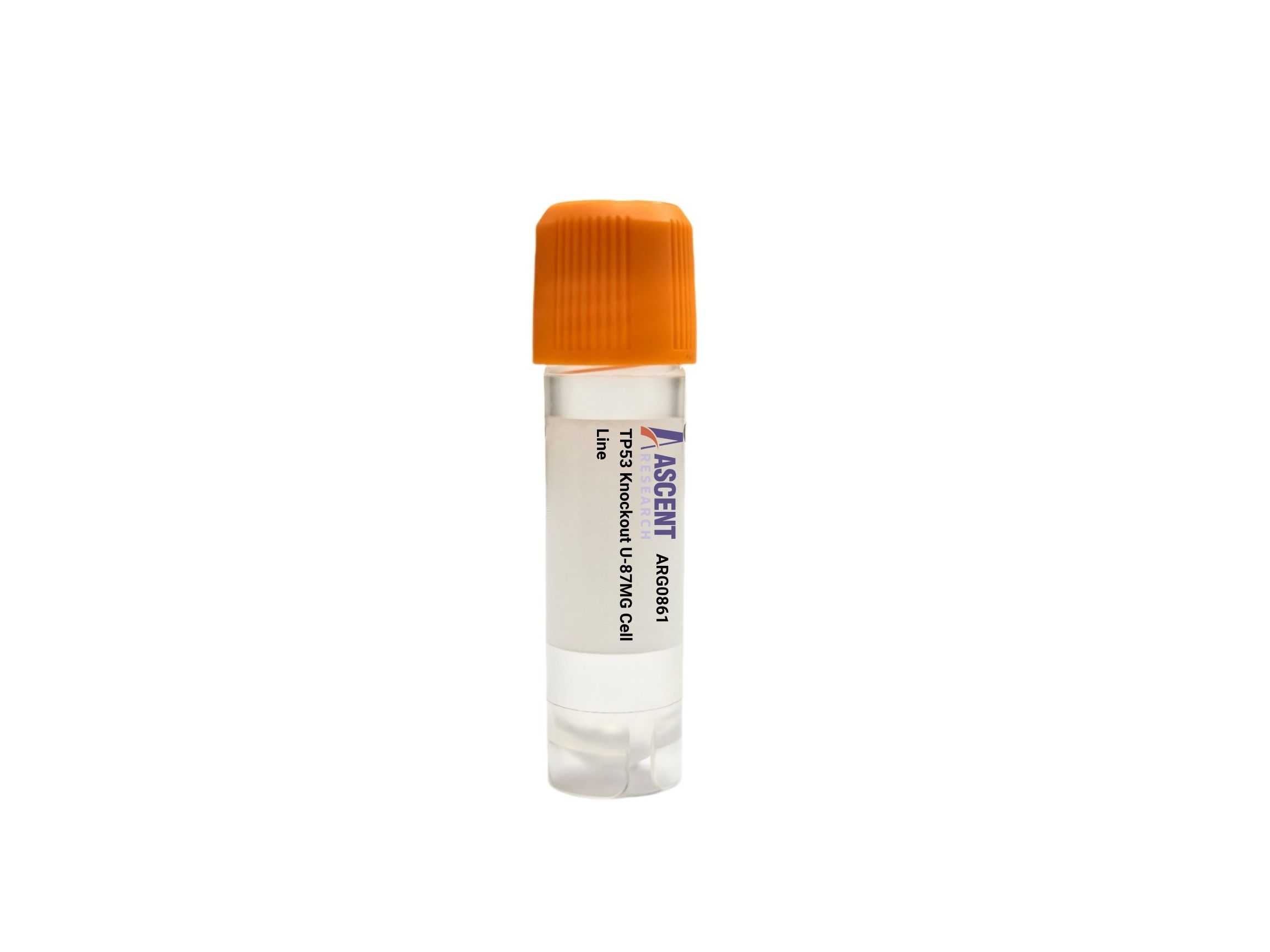
The TP53 Knockout U-87MG Cell Line is a CRISPR/Cas9-edited knockout cell line derived from the U-87MG human glioblastoma cell line, designed to eliminate p53 tumor suppressor function. This loss-of-function model enables precise investigation of p53-dependent processes, as p53 regulates cell cycle arrest, apoptosis, and DNA repair via downstream targets such as CDKN1A (p21) and BAX. Widely applicable in neuro-oncology, this knockout line is ideal for studying chemoresistance in glioblastoma with temozolomide, screening p53-dependent drugs like Nutlin-3, and analyzing DNA damage responses. The model also supports apoptosis assays, cell cycle analysis, and genomic instability assessments, providing a versatile platform for mechanistic and translational research.
The TP53 Knockout U-87MG Cell Line is a CRISPR/Cas9-mediated gene-disrupted derivative of the U-87MG human glioblastoma cell line, engineered to ablate functional p53 protein expression. This product provides a stable, loss-of-function model for investigating the multifaceted roles of the TP53 tumor suppressor gene in a malignant glial context.
The U-87MG host cell line was originally established from a male patient with glioblastoma multiforme and is widely employed as a model system for malignant glioma research. It retains characteristic features of glioblastoma, such as rapid proliferation, invasive potential, and genetic alterations that enable the study of tumor biology. The parental U-87MG line harbors wild-type TP53, establishing a clean background for assaying p53-dependent phenotypes following gene disruption.
p53 functions as a sequence-specific transcription factor that coordinates cellular responses to genotoxic stress, including DNA damage, oncogene activation, and hypoxia. Upstream kinases such as ATM and ATR phosphorylate p53 following DNA damage, while the ARF tumor suppressor inhibits MDM2-mediated degradation, leading to p53 stabilization. Activated p53 transcriptionally induces a broad array of target genes, including CDKN1A (p21) for cell cycle arrest at G1/S and G2/M checkpoints, pro-apoptotic factors BAX, BBC3 (PUMA), and PMAIP1 (NOXA) to initiate mitochondrial apoptosis, and GADD45A for DNA repair. Additionally, p53 upregulates MDM2 in a negative feedback loop, and modulates metabolic and senescence programs through targets like TIGAR and RRM2B. In TP53 knockout cells, this entire regulatory network is disabled.
Loss of p53 function is a hallmark of glioblastoma progression, occurring in a significant fraction of tumors through mutation or alternative mechanisms. In the U-87MG background, TP53 knockout recapitulates a critical oncogenic event, leading to impaired DNA damage checkpoints, enhanced proliferation, and resistance to apoptotic stimuli. This model allows researchers to investigate how p53 deficiency contributes to the aggressive phenotype of glioblastoma, including its notorious chemoresistance to agents such as temozolomide, and to probe synthetic lethal interactions that may be exploited therapeutically.
Researchers can utilize this TP53 knockout cell line in a variety of experimental contexts, including comparative studies of p53-dependent and -independent responses to DNA-damaging agents, high-throughput drug sensitivity screens with agents like Nutlin-3 or temozolomide, and investigation of p53-mediated apoptosis using Annexin V staining. Mechanistic studies can employ qPCR, western blotting, and ChIP-qPCR to examine transcriptional outputs and protein interactions. Further applications include assessing genomic instability via comet assay, monitoring DNA repair kinetics through ??H2AX foci formation, and modeling tumor evolution under p53 deficiency. For detailed technical specifications or to discuss custom projects, please contact Ascent Research.