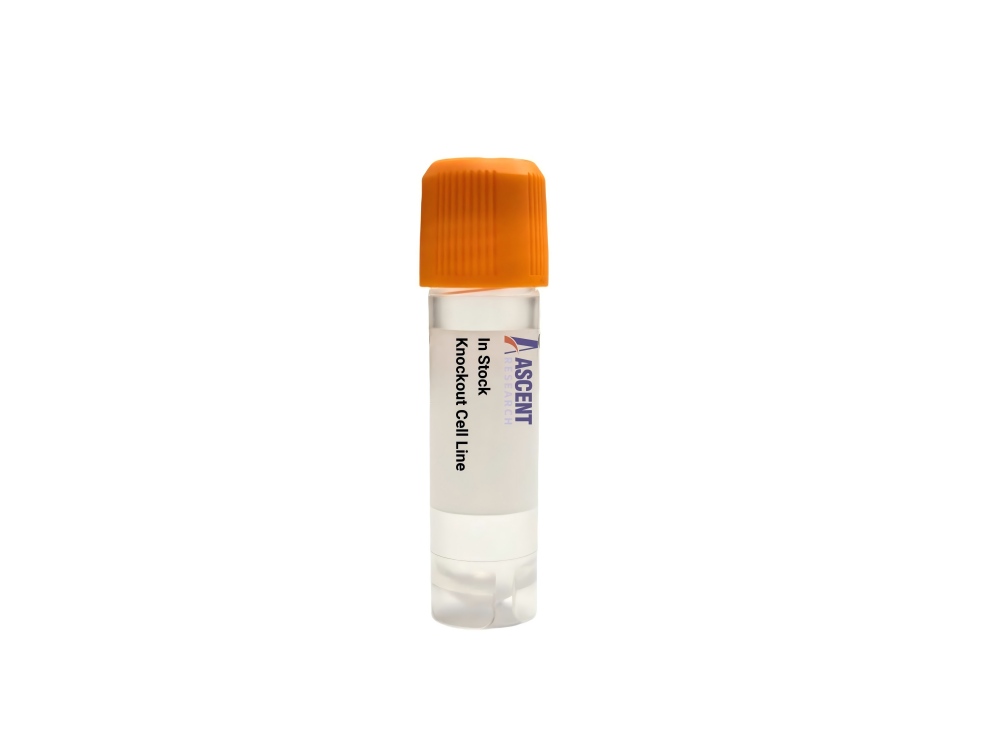
In Stock Cell Lines
Mus musculus (Mouse)
Ascites
Adherent
The Clec16a Knockout RAW 264.7 Cell Line provides a CRISPR/Cas9-edited loss-of-function system in the RAW 264.7 murine macrophage background. Disruption of Clec16a, a regulator of autophagy and mitophagy, allows dissection of its role in mitochondrial quality control and immune signaling. The protein interacts with E3 ubiquitin ligases NRDP1 and Parkin, influencing pathways such as NF-??B and LC3-mediated autophagosome formation. Researchers can employ this model to investigate autophagy mechanisms via western blotting, RT-qPCR, and immunofluorescence, and to study immune dysfunction related to autoimmune disorders like type 1 diabetes and rheumatoid arthritis. Cytokine secretion profiling and mitochondrial mass analysis by flow cytometry are representative downstream assays.
 MYL6B Knockout NCI-H1299 Polyclonal Cells
Cat. No. ARG17478
ERN1 Knockout HEK293 Polyclonal Cells
Cat. No. ARG12664
MYH7 Knockout T47D Polyclonal Cells
Cat. No. ARG12023
IP6K1 Knockout A2780 Polyclonal Cells
Cat. No. ARG29229
DTNB Knockout HAP1 Polyclonal Cells
Cat. No. ARG39896
COMMD1 Knockout HEK293T Polyclonal Cells
Cat. No. ARG3576
MYL6B Knockout NCI-H1299 Polyclonal Cells
Cat. No. ARG17478
ERN1 Knockout HEK293 Polyclonal Cells
Cat. No. ARG12664
MYH7 Knockout T47D Polyclonal Cells
Cat. No. ARG12023
IP6K1 Knockout A2780 Polyclonal Cells
Cat. No. ARG29229
DTNB Knockout HAP1 Polyclonal Cells
Cat. No. ARG39896
COMMD1 Knockout HEK293T Polyclonal Cells
Cat. No. ARG3576
The Clec16a Knockout RAW 264.7 Cell Line is a CRISPR/Cas9-edited knockout cell line derived from the RAW 264.7 murine macrophage model. This product features targeted disruption of the Clec16a gene (Mus musculus), eliminating functional expression of the encoded protein. The knockout cell line serves as a defined loss-of-function system for dissecting Clec16a-dependent mechanisms in macrophage biology and immune regulation. It enables precise investigation of autophagy, mitophagy, and immune signaling pathways without relying on transient gene silencing approaches. Researchers can employ this stable knockout model to examine steady-state and stimulus-induced cellular responses in a well-characterized host background.
The host RAW 264.7 cell line is an Abelson murine leukemia virus-transformed macrophage line originating from BALB/c mice. These cells function as professional immune effector cells, exhibiting robust phagocytic activity, cytokine production, and antigen presentation capabilities. RAW 264.7 cells are widely used to study macrophage activation, inflammatory signaling, and innate immune mechanisms. Their transformed nature permits continuous culture while retaining many primary macrophage features, including responsiveness to toll-like receptor ligands and inflammasome activators. This background provides a physiologically relevant context for exploring Clec16a function in immune homeostasis.
Clec16a regulates autophagy and mitophagy through interaction with E3 ubiquitin ligases such as NRDP1 and Parkin, forming complexes that promote ubiquitin-dependent mitochondrial clearance and autophagosome formation. Its activity is modulated by nutrient deprivation, mitochondrial stress, and inflammatory cytokines, and it governs downstream processes including LC3-mediated autophagosome maturation and NF-??B signaling. Clec16a operates within the PINK1/Parkin mitophagy axis, interacting with NRDP1 and ubiquitin to coordinate removal of damaged mitochondria. Disruption of this gene impairs mitochondrial quality control and alters immune effector functions, highlighting its role at the interface of cellular metabolism and immunity.
In RAW 264.7 macrophages, Clec16a knockout induces mitophagy defects and may lead to accumulation of dysfunctional mitochondria, altered ROS production, and skewed cytokine profiles. These phenotypes mirror aspects of autoimmune disorders such as type 1 diabetes, multiple sclerosis, and rheumatoid arthritis, where Clec16a variants are linked to disease risk. The model thus enables dissection of how autophagy failure contributes to immune dysregulation in a disease-relevant cell type.
Applications include autophagy analysis via immunoblotting for LC3-II/p62, RT-qPCR of autophagy genes, and immunofluorescence for autophagosomes. Mitochondrial mass can be quantified by flow cytometry, and immune function assessed through cytokine secretion profiling. The cell line supports examination of Clec16a-dependent signaling under various stimuli. For further details, contact Ascent Research.