In Stock Cell Lines
Homo sapiens (Human)
Uterus (cervix)
Adherent
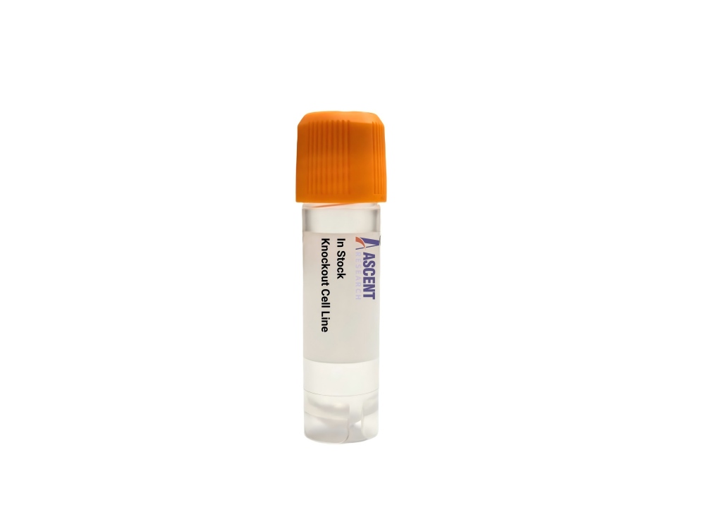
The ATM Knockout HeLa Cell Line is a CRISPR/Cas9-edited human knockout cell line derived from HeLa cervical adenocarcinoma cells, designed for studying ATM serine/threonine kinase function in DNA damage signaling. ATM is activated by DNA double-strand breaks via the MRN complex and phosphorylates downstream targets such as p53 and CHK2 to control cell cycle checkpoints, DNA repair, and apoptosis. This model is particularly valuable for investigating p53-independent DNA damage responses, given HeLa's HPV18-driven p53 inactivation. Applications include DNA repair analysis by ??-H2AX foci formation, clonogenic survival assays for chemosensitivity, and cell cycle profiling, supporting cancer biology and radiobiology research.
 HCLS1 Knockout K562 Polyclonal Cells
Cat. No. ARG20531
PDPN Knockout 143B Polyclonal Cells
Cat. No. ARG11812
AKT1S1 Knockout 786-O Polyclonal Cells
Cat. No. ARG24874
ITFG1 Knockout Hela Polyclonal Cells
Cat. No. ARG26286
Me1 Knockout HEK293T Polyclonal Cells
Cat. No. ARG4538
MPPED2 Knockout HEK293T Polyclonal Cells
Cat. No. ARG4533
HCLS1 Knockout K562 Polyclonal Cells
Cat. No. ARG20531
PDPN Knockout 143B Polyclonal Cells
Cat. No. ARG11812
AKT1S1 Knockout 786-O Polyclonal Cells
Cat. No. ARG24874
ITFG1 Knockout Hela Polyclonal Cells
Cat. No. ARG26286
Me1 Knockout HEK293T Polyclonal Cells
Cat. No. ARG4538
MPPED2 Knockout HEK293T Polyclonal Cells
Cat. No. ARG4533
The ATM Knockout HeLa Cell Line is a CRISPR/Cas9-edited human cell line derived from the HeLa cervical adenocarcinoma line, featuring targeted disruption of the ATM (ataxia telangiectasia mutated) gene to create a loss-of-function model for studying ATM-dependent processes. It is designed to facilitate mechanistic investigations into DNA damage response, cell cycle regulation, and apoptosis pathways in a well-characterized epithelial carcinoma context.
HeLa cells are an immortalized human epithelial cell line originally derived from a cervical adenocarcinoma and are widely employed in cancer research, virology, and DNA damage signaling studies. They harbor human papillomavirus type 18 (HPV18) sequences, which contribute to the degradation of p53 via the E6 oncoprotein, rendering the cells functionally p53-deficient under normal conditions. This unique genetic background provides a distinct context for investigating ATM-driven responses that are independent of the canonical p53 pathway, and the cells?? robust growth and adaptability make them a standard platform for cell-based assays in molecular oncology.
ATM functions as a central serine/threonine kinase in the DNA damage response, activated by DNA double-strand breaks (DSBs) through the MRE11-RAD50-NBS1 (MRN) complex. It phosphorylates numerous downstream targets, including p53 (TP53), CHK2 (CHEK2), and H2AX (H2AFX). These events mediate cell cycle arrest via the ATM-p53-p21 and ATM-CHK2-CDC25 axes, promote DNA repair via homologous recombination and non-homologous end joining, or trigger apoptosis. ATM interacts with ATR, DNA-PKcs (PRKDC), 53BP1 (TP53BP1), and MDC1 to coordinate repair and maintain genomic stability. Its loss results in defective checkpoint control and genomic instability, hallmarks of cancer predisposition.
In the HeLa background, where p53 function is abrogated by HPV18 E6, ATM knockout enables the dissection of p53-independent DNA damage signaling pathways. This cell line allows researchers to examine how ATM modulates cell cycle checkpoints, repair factor recruitment, and survival decisions without the confounding influence of p53-mediated transcriptional responses. The resulting genomic instability and altered sensitivity to DNA-damaging agents mirror aspects of ATM-deficient tumors, making this model highly relevant for studying cancers driven by ATM loss or mutations, such as certain breast cancers, lymphomas, and leukemias.
The ATM Knockout HeLa Cell Line is suited for diverse applications, including DNA damage response characterization via Western blotting for ??-H2AX and immunofluorescence detection of ??-H2AX foci. The comet assay and clonogenic survival assays assess genomic integrity and chemosensitivity, while flow cytometry reveals cell cycle checkpoint defects. RT-qPCR for p21 and other targets further profiles pathway activity. This knockout cell line supports radiation biology, chemosensitivity testing, and genome stability studies. For additional information, please contact Ascent Research.