Genome-edited Cells
Unknown
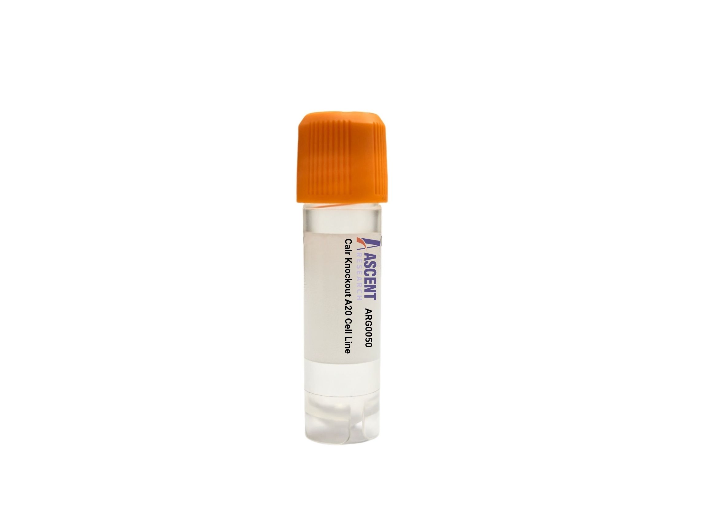
Calr Knockout A20 is a CRISPR/Cas9-edited mouse B cell lymphoma cell line with disruption of the ER chaperone gene Calr. In the BALB/c-derived A20 antigen-presenting B lymphocyte background, loss of calreticulin supports studies of ER protein folding, Ca2+ homeostasis, MHC class I assembly, and stress signaling mediated by factors such as CALNEXIN, PDIA3, ATF6, XBP1, and PERK/EIF2AK3. This model is well suited for analysis of lymphoma biology, cancer immunology, unfolded protein response regulation, antigen presentation, immunogenic cell death, apoptosis, and proteostasis using flow cytometry, western blotting, RNA-seq, and ER stress assays.
 NUDT15 Knockout jurkat Polyclonal Cells
Cat. No. ARG13317
ABCC1 Knockout HGC-27 Polyclonal Cells
Cat. No. ARG29361
BCAR1 Knockout A549 Polyclonal Cells
Cat. No. ARG31922
CAB39 Knockout MES-OV Polyclonal Cells
Cat. No. ARG41702
NRAS Knockout 786-O Polyclonal Cells
Cat. No. ARG4688
GLUD1 Knockout AGS Polyclonal Cells
Cat. No. ARG2534
NUDT15 Knockout jurkat Polyclonal Cells
Cat. No. ARG13317
ABCC1 Knockout HGC-27 Polyclonal Cells
Cat. No. ARG29361
BCAR1 Knockout A549 Polyclonal Cells
Cat. No. ARG31922
CAB39 Knockout MES-OV Polyclonal Cells
Cat. No. ARG41702
NRAS Knockout 786-O Polyclonal Cells
Cat. No. ARG4688
GLUD1 Knockout AGS Polyclonal Cells
Cat. No. ARG2534
The Calr Knockout A20 Cell Line is a CRISPR/Cas9-engineered murine B cell lymphoma model in which the Calr gene has been disrupted to eliminate functional calreticulin expression. This stable in vitro knockout system is generated in A20 cells, a mouse BALB/c-derived antigen-presenting B lymphocyte line that expresses immunoglobulin and exhibits cytokine-responsive signaling behavior. The model is designed for mechanistic studies of endoplasmic reticulum proteostasis, calcium handling, antigen presentation, and stress-response pathways in a hematologic cancer cell context.
A20 cells are widely used as an experimental model for B cell receptor-associated signaling, antigen processing and presentation, apoptosis regulation, and NF-kB-dependent immune processes. As a murine lymphoma line with established relevance to B cell biology and inflammatory signaling, A20 provides a tractable host for dissecting immune-regulatory pathways under basal and stimulated conditions. Its utility in studies of cytokine responses, survival signaling, and lymphoma-associated phenotypes makes it particularly suitable for evaluating how ER-resident chaperone systems influence immune cell function and stress adaptation.
CALR encodes an ER luminal lectin-like chaperone that functions within the calnexin-calreticulin cycle to promote folding and quality control of nascent glycoproteins while buffering ER Ca2+ stores. CALR interacts with CALNEXIN, PDIA3/ERp57, TAPBP/tapasin, TAP1, TAP2, beta-2-microglobulin, and MHC class I heavy chains to support peptide loading and efficient MHC class I assembly. Its expression and activity are regulated by ER stress-associated pathways involving ATF6, IRE1/ERN1-XBP1, and PERK/EIF2AK3-eIF2alpha-ATF4-DDIT3/CHOP signaling, particularly under disturbed ER Ca2+ homeostasis or proteotoxic stress. Loss of Calr is therefore expected to alter ER quality control, unfolded protein response output, properly folded glycoprotein recovery, MHC class I peptide loading efficiency, HSPA5/BiP and DDIT3/CHOP induction during stress, ER Ca2+-dependent apoptotic responses, and stress-induced cell-surface calreticulin exposure.
In A20 lymphoma cells, Calr deficiency provides a relevant platform for linking ER chaperone loss to B cell-specific immune phenotypes. Because A20 cells integrate antigen presentation capacity with cytokine-responsive and NF-kB-regulated programs, this knockout can be used to examine how defective ER folding machinery reshapes immune surface phenotype, stress tolerance, and lymphoma cell adaptation. The model is pertinent to studies of lymphoma biology, cancer immunology, ER stress-related disorders, autoimmunity, and broader mechanisms relevant to hematologic malignancies and myeloproliferative disease research.
Researchers can apply this cell line in western blotting, RT-qPCR, and RNA-seq workflows to quantify UPR pathway components such as Hspa5, Xbp1 target genes, and Ddit3. Flow cytometry can be used to assess surface MHC class I expression, peptide presentation-related phenotypes, and stress-induced calreticulin externalization. Additional applications include immunofluorescence for ER morphology or protein localization, co-immunoprecipitation of CALR-associated folding complexes, ER stress induction assays, phospho-signaling analysis of PERK pathway activation, Ca2+ flux measurements, apoptosis assays, antigen presentation studies, and proteostasis or drug-sensitivity profiling in response to ER stressors or immune-modulatory compounds. Researchers may contact Ascent Research for additional technical information, product details, or related gene-edited cell models.