In Stock Cell Lines
Homo sapiens (Human)
Kidney
Adherent
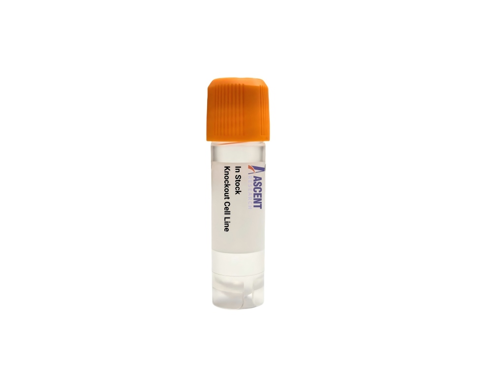
The FUNDC1 Knockout HEK293T Cell Line is a CRISPR/Cas9-edited cell line lacking expression of the FUNDC1 mitophagy receptor, based on the high-transfection human embryonic kidney HEK293T host. It enables precise dissection of hypoxia-induced mitochondrial autophagy, where FUNDC1 dephosphorylation by PGAM5 facilitates LC3 binding and mitochondrial recruitment. Key regulators include HIF-1??, CK2, and Src kinases. This knockout line supports research into cancer, neurodegeneration, and ischemia-reperfusion injury using techniques such as immunoblotting, fluorescence colocalization, and mitophagy flux assays. It is a robust system for validating therapeutic targets and exploring mitochondrial quality control mechanisms. Contact Ascent Research for further details.
 NME7 Knockout A2780 Polyclonal Cells
Cat. No. ARG18518
MAN2C1 Knockout A549 Polyclonal Cells
Cat. No. ARG10512
DCXR Knockout Hela Polyclonal Cells
Cat. No. ARG7937
HMOX1 Knockout jurkat Polyclonal Cells
Cat. No. ARG34266
APOBEC3A Knockout HT29 Polyclonal Cells
Cat. No. ARG36158
MSRA Knockout AGS Polyclonal Cells
Cat. No. ARG2245
NME7 Knockout A2780 Polyclonal Cells
Cat. No. ARG18518
MAN2C1 Knockout A549 Polyclonal Cells
Cat. No. ARG10512
DCXR Knockout Hela Polyclonal Cells
Cat. No. ARG7937
HMOX1 Knockout jurkat Polyclonal Cells
Cat. No. ARG34266
APOBEC3A Knockout HT29 Polyclonal Cells
Cat. No. ARG36158
MSRA Knockout AGS Polyclonal Cells
Cat. No. ARG2245
The FUNDC1 Knockout HEK293T Cell Line is a CRISPR/Cas9-edited human cell line with targeted disruption of the FUNDC1 gene, eliminating expression of the mitophagy receptor. This knockout model provides a stable loss-of-function system for dissecting FUNDC1-dependent mitochondrial quality control and hypoxia-induced autophagy.
The host HEK293T cell line, derived from human embryonic kidney epithelium, stably expresses SV40 large T antigen, enhancing episomal plasmid replication and recombinant protein expression. Their high transfection efficiency and robust growth make them a preferred host for gene function and signaling studies.
FUNDC1 localizes to the mitochondrial outer membrane, where it acts as a hypoxia-responsive mitophagy receptor. Under low oxygen, HIF-1?? upregulates FUNDC1, and PGAM5 dephosphorylates it at Ser13, promoting interaction with LC3B and GABARAP to drive autophagosomal recruitment. Normoxic conditions see CK2 and Src-mediated phosphorylation that inhibit LC3 binding, while BCL2L1 binding suppresses mitophagy. FUNDC1 also interfaces with mitochondrial dynamics proteins DRP1 and OPA1, the E3 ligase MARCH5, and kinase ULK1.
In the HEK293T background, this knockout cell line provides a clean system to dissect the molecular steps of hypoxia-induced mitophagy, free from endogenous FUNDC1 activity. HEK293T cells are responsive to hypoxic stimuli and are extensively used in signaling and metabolic studies, making this line particularly suitable for unraveling FUNDC1??s role in mitochondrial stress responses and cell survival. It is valuable for exploring how FUNDC1 integrates signals to control mitochondrial turnover, relevant to pathologies such as neurodegeneration, cancer, and ischemia-reperfusion injury.
Typical assays include immunoblotting to monitor FUNDC1 loss and LC3 lipidation, immunofluorescence for quantification of LC3 puncta colocalizing with mitochondrial markers, and flow cytometry using mt-Keima or mito-QC reporters to measure mitophagy flux. Seahorse-based mitochondrial stress tests and co-immunoprecipitation of the FUNDC1-LC3 complex further enable rigorous functional analyses. This knockout line is thus a powerful tool for target validation in drug discovery, mechanistic studies of mitochondrial quality control, and investigation of FUNDC1-associated signaling networks. For further technical details or custom applications, please contact Ascent Research.