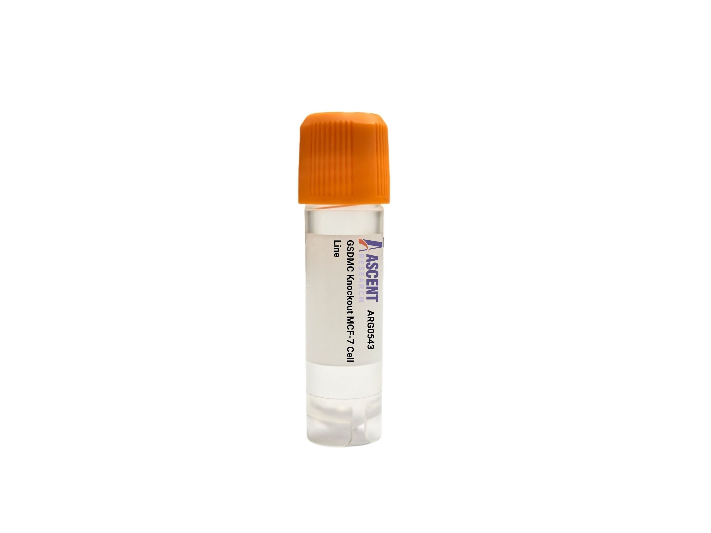
Genome-edited Cells
Breast (mammary gland)
The GSDMC Knockout MCF-7 Cell Line is a CRISPR/Cas9-edited human cell line lacking functional GSDMC, a pyroptosis executioner that forms membrane pores upon cleavage by caspase-8 downstream of TNF-alpha/TNFR1 signaling. Derived from the ER+ MCF-7 breast adenocarcinoma line, this model enables dissection of inflammatory cell death mechanisms in a hormone-responsive cancer background. It is ideal for investigating pyroptosis induction, IL-1beta and IL-18 release, and drug sensitivity. Applications include LDH release assays, ELISA, flow cytometry, and xenograft studies, offering insights into TNF-alpha-driven cell death and breast cancer biology. Contact Ascent Research for more information.
 CIB1 Knockout SK-HEP-1 Polyclonal Cells
Cat. No. ARG15213
CIB1 Knockout SK-HEP-1 Polyclonal Cells
Cat. No. ARG15213
 Grk5 Knockout RAW 264.7 Cell Line
Cat. No. ARG43889
GPT2 Knockout HGC-27 Polyclonal Cells
Cat. No. ARG29864
Grk5 Knockout RAW 264.7 Cell Line
Cat. No. ARG43889
GPT2 Knockout HGC-27 Polyclonal Cells
Cat. No. ARG29864
 Rabbit Prostate Fibroblasts
Cat. No. ARP0747
Rabbit Prostate Fibroblasts
Cat. No. ARP0747
 Mouse Renal Artery Smooth Muscle Cells
Cat. No. ARP0457
Mouse Renal Artery Smooth Muscle Cells
Cat. No. ARP0457
 Tmem176b Knockout EL4 Cell Line
Cat. No. ARG0230
Tmem176b Knockout EL4 Cell Line
Cat. No. ARG0230
The GSDMC Knockout MCF-7 Cell Line is a CRISPR/Cas9-edited human cell line that carries a targeted disruption of the GSDMC gene, resulting in a loss-of-function model for studying pyroptosis and inflammatory cell death. MCF-7 cells, derived from a metastatic mammary adenocarcinoma, serve as a well-established estrogen receptor-positive (ER+) breast cancer model. This knockout cell line provides a genetically defined system to dissect the molecular mechanisms governing GSDMC-mediated pore formation and downstream cytokine release.
MCF-7 cells are adherent epithelial cells originally isolated from the pleural effusion of a patient with metastatic breast adenocarcinoma. They retain expression of estrogen receptor alpha (ER??) and are widely used to model luminal A breast cancer, hormone responsiveness, and metastatic progression. The cells exhibit typical epithelial morphology and are sensitive to hormonal and chemotherapeutic agents, making them a versatile platform for both fundamental and translational oncology research.
GSDMC encodes a member of the gasdermin family that functions as a key executioner of pyroptosis, a lytic and highly inflammatory form of programmed cell death. Upon activation of the TNF-alpha/TNFR1 signaling axis, caspase-8 cleaves GSDMC, liberating its N-terminal domain. This fragment then translocates to and oligomerizes within the plasma membrane, forming pores that disrupt cellular ionic homeostasis, drive osmotic swelling, and ultimately cause membrane rupture. The process culminates in the extracellular release of pro-inflammatory cytokines such as IL-1beta and IL-18, along with alarmins including HMGB1 and LDH. Upstream regulators such as TGF-beta, STAT3, NF-kB, and IL-1beta modulate GSDMC expression and activity, placing the protein at a nexus of cell death and inflammatory signaling. Interaction with caspase-1 and caspase-4 further diversifies its activation landscape.
In breast cancer, particularly the ER+ subtype represented by MCF-7 cells, GSDMC-mediated pyroptosis shapes the tumor microenvironment and therapeutic response. Loss of GSDMC function may alter sensitivity to TNF-alpha-mediated cytotoxicity and influence the immunogenicity of dying cells. This knockout model enables dissection of how GSDMC-dependent pore formation affects drug-induced cell death, cytokine release profiles, and the balance between apoptosis and pyroptosis, with potential implications for tumor progression and metastasis.
Typical research applications include quantifying pyroptosis by flow cytometry and LDH release, measuring IL-1beta and IL-18 secretion via ELISA, and assessing caspase-8 activity following TNF-alpha stimulation. The cell line is also suitable for Western blot and RT-qPCR to confirm GSDMC disruption and for functional assays such as MTT cell viability, Transwell migration/invasion, and drug sensitivity testing with doxorubicin or TNF-alpha. Immunofluorescence visualization of GSDMC-N pore complexes and tumor xenograft studies further expand its utility. For further technical details or to discuss experimental applications, please contact Ascent Research.