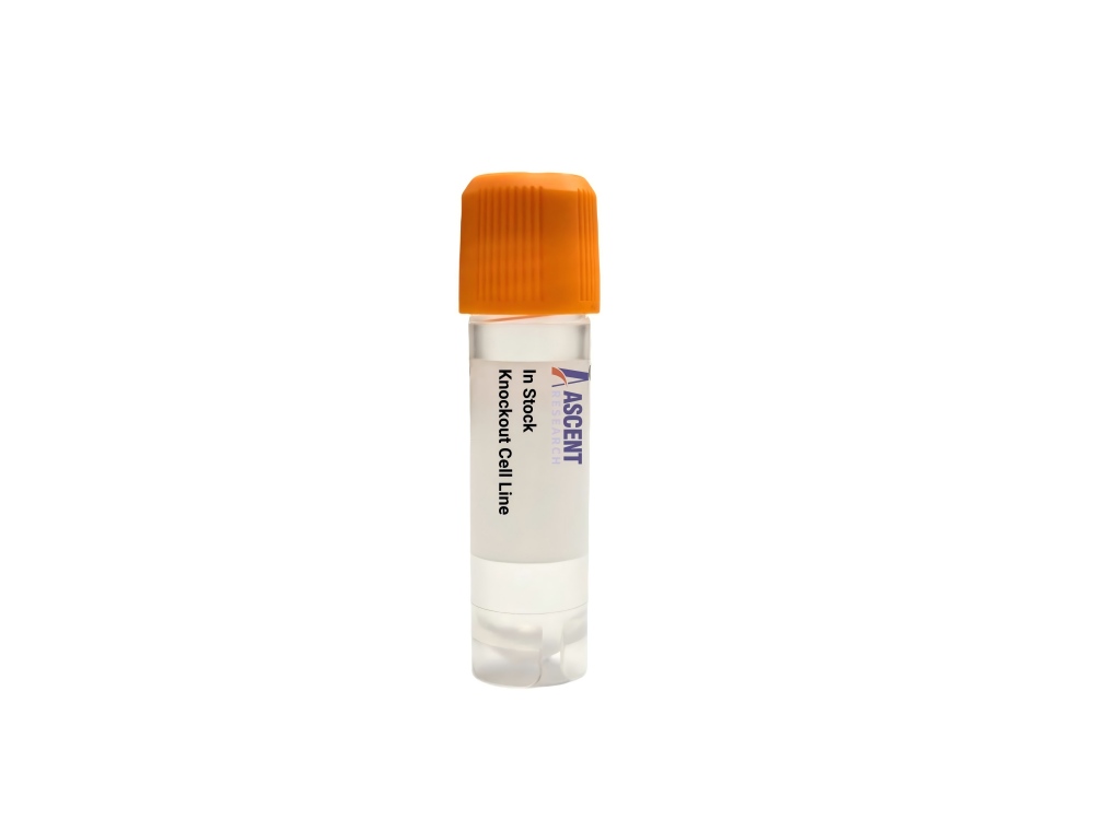
In Stock Cell Lines
The Hmgb1 Knockout iBMDM Cell Line is a CRISPR/Cas9-edited knockout cell line derived from immortalized mouse bone marrow-derived macrophages (iBMDMs) that lacks the alarmin HMGB1. This model abrogates HMGB1-mediated activation of TLR4 and RAGE pathways, disrupting downstream NF-??B and MAPK signaling and attenuating pro-inflammatory cytokine production. It serves as a critical tool for dissecting DAMP-driven innate immunity in sepsis, acute lung injury, rheumatoid arthritis, and cancer research. Applications include western blotting, ELISA, NF-??B luciferase reporter assays, cytokine RT-qPCR, and functional studies such as phagocytosis and migration assays.
 GPR173 Knockout HAP1 Polyclonal Cells
Cat. No. ARG22329
ETFB Knockout K562 Polyclonal Cells
Cat. No. ARG19477
GNS Knockout A2780 Polyclonal Cells
Cat. No. ARG29014
CBL Knockout NCI-H1299 Polyclonal Cells
Cat. No. ARG42666
ESRRA Knockout Hela Polyclonal Cells
Cat. No. ARG7851
GPR173 Knockout HAP1 Polyclonal Cells
Cat. No. ARG22329
ETFB Knockout K562 Polyclonal Cells
Cat. No. ARG19477
GNS Knockout A2780 Polyclonal Cells
Cat. No. ARG29014
CBL Knockout NCI-H1299 Polyclonal Cells
Cat. No. ARG42666
ESRRA Knockout Hela Polyclonal Cells
Cat. No. ARG7851
 Rabbit Thyroid Fibroblasts
Cat. No. ARP0758
Rabbit Thyroid Fibroblasts
Cat. No. ARP0758
The Hmgb1 Knockout iBMDM Cell Line is a CRISPR/Cas9-edited knockout cell line derived from immortalized C57BL/6 mouse bone marrow-derived macrophages (iBMDMs) carrying a targeted disruption of the Hmgb1 gene. This loss-of-function model enables precise investigation of HMGB1-dependent signaling in a well-characterized macrophage background.
The parental iBMDM cell line originates from C57BL/6 mouse bone marrow-derived macrophages that have been immortalized, retaining key macrophage functions including phagocytosis, antigen presentation, and cytokine secretion. This homogeneous and genetically tractable host system provides a robust platform for dissecting macrophage-specific gene functions and signaling pathways.
HMGB1 is a highly conserved DNA-binding protein that functions as a damage-associated molecular pattern (DAMP) when released into the extracellular milieu during cellular stress or necrotic cell death. Extracellular HMGB1 engages receptors TLR4 and RAGE, triggering MyD88-dependent signal transduction that culminates in the activation of NF-??B and MAP kinase cascades, including ERK1/2 and p38. This signaling axis drives the transcriptional upregulation of pro-inflammatory mediators such as IL-6 and TNF-??. Release of HMGB1 is stimulated by upstream signals including TNF-??, IL-1??, lipopolysaccharide (LPS), and oxidative stress, and it interacts directly with p53 and the NF-??B p65 (RELA) subunit within the nucleus, linking inflammation to transcriptional and cell fate decisions.
Ablation of Hmgb1 in iBMDM cells abrogates the major alarmin-driven feedforward loop that amplifies macrophage inflammatory responses. In the knockout cells, stimulation with LPS, TNF-??, or other danger signals fails to fully activate TLR4/RAGE-dependent NF-??B and MAPK pathways, resulting in attenuated secretion of IL-6, TNF-??, and other effector cytokines. Consequently, key macrophage functional outputs??including phagocytic capacity, antigen presentation, and chemotactic migration??are altered, making this cell line a definitive tool for distinguishing HMGB1-dependent from HMGB1-independent mechanisms in macrophage biology.
Researchers employ this knockout cell line in a variety of experimental settings to probe innate immune regulation. It is suitable for DAMP signaling analysis, where the absence of HMGB1 clarifies its contribution to TLR4/RAGE-mediated inflammatory cascades; for sepsis and acute lung injury models, where HMGB1 acts as a late mediator; and for rheumatoid arthritis and ischemia-reperfusion injury studies, where extracellular HMGB1 exacerbates tissue damage. The line is also used in macrophage polarization experiments to determine how HMGB1 influences M1/M2 balance, and in high-throughput screening campaigns aimed at identifying small-molecule HMGB1 inhibitors. Representative assay readouts include HMGB1 western blotting and ELISA to confirm knockout, NF-??B luciferase reporters for pathway activity, RT-qPCR for cytokine transcripts, immunofluorescence for subcellular localization, and functional assays such as phagocytosis and transwell migration. For further technical details, please contact Ascent Research.