Genome-edited Cells
Bone
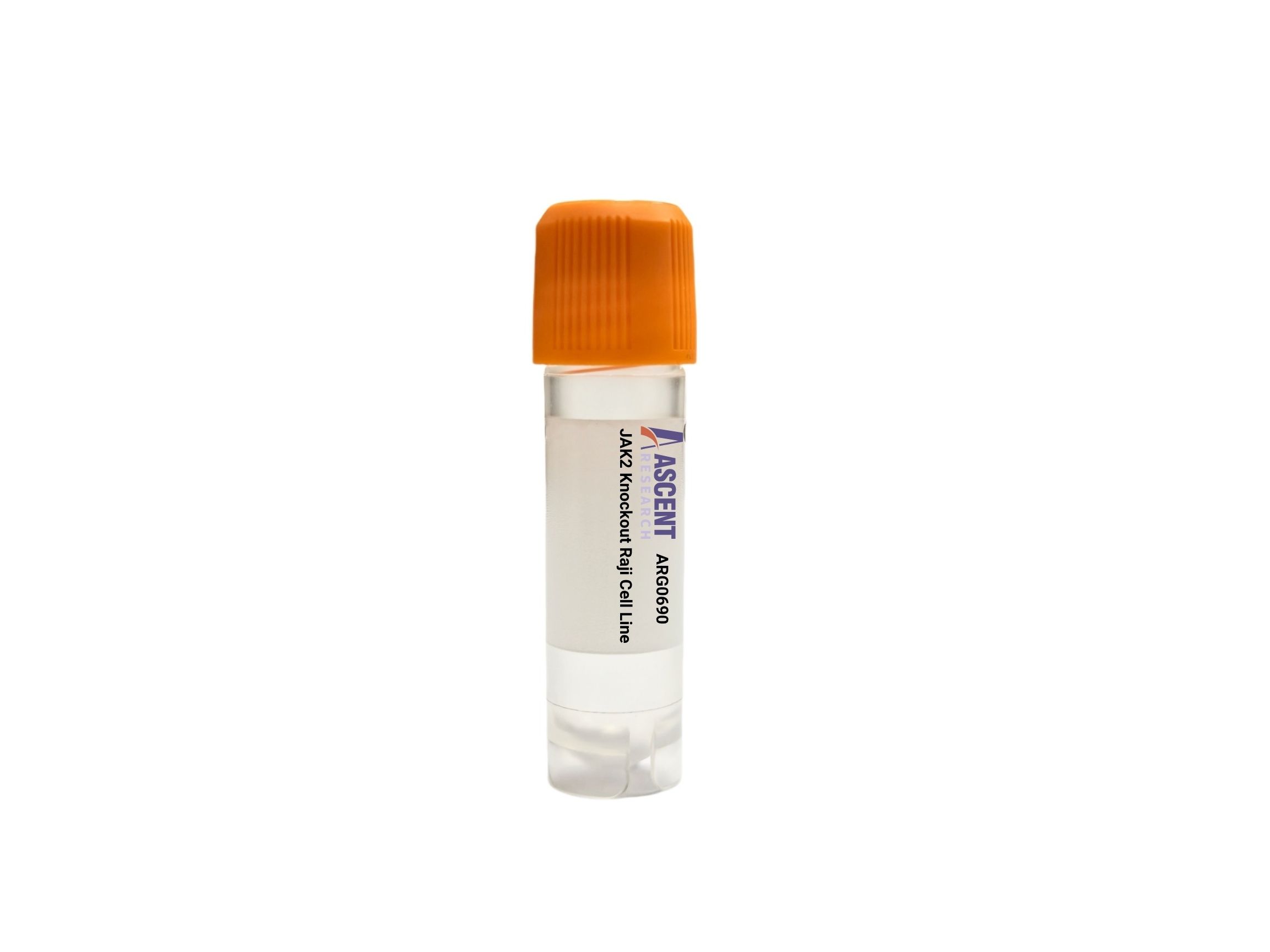
The JAK2 Knockout Raji Cell Line is a CRISPR/Cas9-edited knockout model derived from the human Raji B lymphocyte line, a Burkitt lymphoma-derived cell system. JAK2 is a non-receptor tyrosine kinase that mediates signaling downstream of cytokine receptors (e.g., EPO, TPO, GM-CSF, IL-3) through the JAK-STAT pathway, regulating critical effectors such as STAT3, STAT5, BCL2L1, and CCND1 in lymphoproliferation and survival. This knockout cell line enables dissection of JAK2-dependent signaling in B-cell malignancy, screening of JAK2 inhibitors like ruxolitinib, and assessment of proliferation, apoptosis, and pathway activation. Representative assays include phospho-STAT5 Western blot, CCK-8 viability, and Annexin V flow cytometry.
 ARSH Knockout HAP1 Polyclonal Cells
Cat. No. ARG21912
GORASP1 Knockout K562 Polyclonal Cells
Cat. No. ARG20479
OCIAD1 Knockout NCI-H1975 Polyclonal Cells
Cat. No. ARG17099
ACOT2 Knockout 786-O Polyclonal Cells
Cat. No. ARG24804
OFD1 Knockout MES-OV Polyclonal Cells
Cat. No. ARG6860
FN3KRP Knockout 786-O Polyclonal Cells
Cat. No. ARG5158
ARSH Knockout HAP1 Polyclonal Cells
Cat. No. ARG21912
GORASP1 Knockout K562 Polyclonal Cells
Cat. No. ARG20479
OCIAD1 Knockout NCI-H1975 Polyclonal Cells
Cat. No. ARG17099
ACOT2 Knockout 786-O Polyclonal Cells
Cat. No. ARG24804
OFD1 Knockout MES-OV Polyclonal Cells
Cat. No. ARG6860
FN3KRP Knockout 786-O Polyclonal Cells
Cat. No. ARG5158
The JAK2 Knockout Raji Cell Line is a CRISPR/Cas9-edited knockout cell line derived from the human Raji B lymphocyte line, providing a defined loss-of-function model for investigating JAK2-dependent signaling pathways. This product features targeted disruption of the JAK2 gene, enabling researchers to dissect the role of JAK2 in cytokine receptor-mediated signal transduction within a neoplastic B-cell environment.
The Raji host cell line is an Epstein-Barr virus (EBV)-positive Burkitt lymphoma-derived B lymphocyte line, widely used as a model for B-cell malignancies. Raji cells retain antigen-presenting capabilities and produce immunoglobulins, making them a relevant system for studying B-cell biology and lymphomagenesis. The transformed phenotype of Raji cells provides a robust platform for examining oncogenic signaling networks.
JAK2 encodes a non-receptor tyrosine kinase that acts downstream of cytokine receptors such as EPOR, MPL, and receptors for GM-CSF, IL-3, and IL-5. Upon ligand binding, JAK2 autophosphorylates and phosphorylates receptor tyrosines, creating docking sites for STAT3, STAT5A, and STAT5B. Phosphorylated STATs dimerize, translocate to the nucleus, and transcriptionally activate pro-proliferative (CCND1, PIM1) and anti-apoptotic (BCL2L1) genes. JAK2 signaling also engages PI3K-AKT-mTOR and MAPK/ERK cascades through adaptor-mediated interactions. The kinase is regulated by SH2B3, SOCS1, SOCS3, and PTPN11, and forms heterodimers with JAK1 or TYK2 in receptor complexes.
In Raji B lymphocytes, JAK2 plays a critical role in transmitting proliferative and survival signals downstream of cytokines that influence B-cell development and malignant transformation. Knockout of JAK2 in this Burkitt lymphoma model abrogates canonical JAK-STAT signaling, leading to impaired cytokine-induced growth and increased susceptibility to apoptosis. This cell line thus represents a disease-relevant tool to study the dependency of neoplastic B cells on JAK2-mediated signal transduction and to evaluate therapeutic strategies targeting the JAK-STAT axis in lymphomas.
Key applications include JAK2 inhibitor screening (e.g., ruxolitinib), cytokine signaling dissection (IL-3, IL-6 stimulation), and functional assays for proliferation (MTT/CCK-8) and apoptosis (Annexin V). Researchers can assess pathway activation via Western blot (phospho-STAT3/5), cell cycle by propidium iodide, and gene expression by RT-qPCR (BCL2L1, CCND1). Co-immunoprecipitation enables study of JAK2?Creceptor interactions. This model supports B-cell lymphoma pathogenesis research and myeloproliferative neoplasm drug evaluation. For further information or technical support, please contact Ascent Research.