Genome-edited Cells
Blood (peripheral blood)
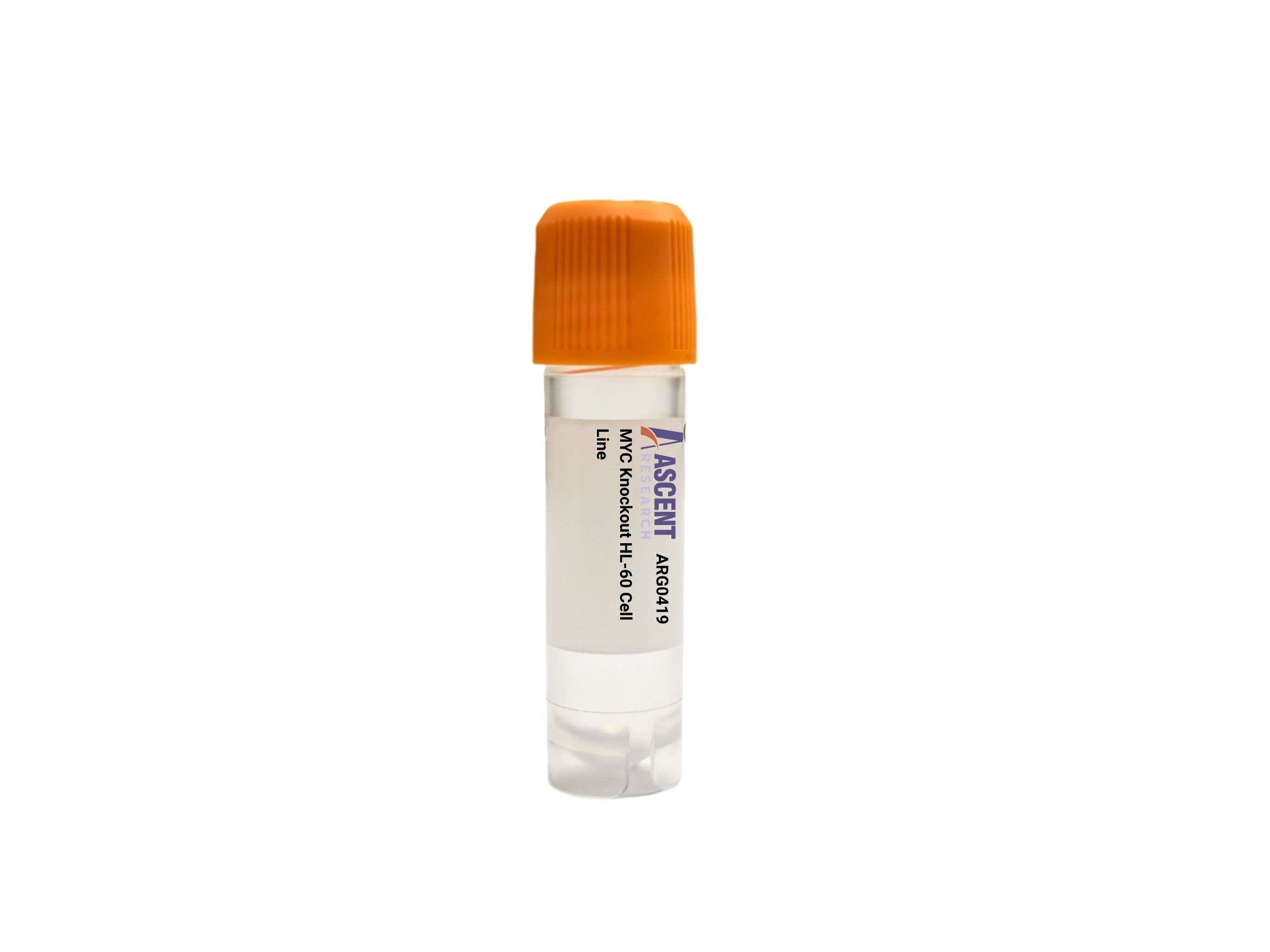
The MYC Knockout HL-60 Cell Line is a CRISPR/Cas9-edited knockout cell model in human acute promyelocytic leukemia HL-60 cells, targeting the MYC transcription factor proto-oncogene. MYC heterodimerizes with MAX to regulate downstream targets such as CCND1 and CDK4, controlling cell cycle progression and metabolism. This p53-null, differentiation-competent line enables precise investigation of MYC-dependent signaling pathways, including Wnt/??-catenin and PI3K/AKT/mTOR cascades. Researchers can employ this knockout to study MYC??s role in leukemic proliferation, apoptosis, and differentiation, as well as to screen pathway inhibitors. The HL-60 background supports functional assays under proliferating and differentiation-inducing conditions, providing a robust platform for cancer biology and drug discovery studies.
 FDX1 Knockout NCI-H1299 Polyclonal Cells
Cat. No. ARG17982
CRADD Knockout jurkat Polyclonal Cells
Cat. No. ARG13002
KEAP1 Knockout Jurkat Polyclonal Cells
Cat. No. ARG23331
GPS2 Knockout MES-OV Polyclonal Cells
Cat. No. ARG24469
FDX1 Knockout NCI-H1299 Polyclonal Cells
Cat. No. ARG17982
CRADD Knockout jurkat Polyclonal Cells
Cat. No. ARG13002
KEAP1 Knockout Jurkat Polyclonal Cells
Cat. No. ARG23331
GPS2 Knockout MES-OV Polyclonal Cells
Cat. No. ARG24469
 CASP1 Knockout HP-1 Cell Line
Cat. No. ARG0424
CASP1 Knockout HP-1 Cell Line
Cat. No. ARG0424
 Aldob Knockout Hepa 1-6 Cell Line
Cat. No. ARG0369
Aldob Knockout Hepa 1-6 Cell Line
Cat. No. ARG0369
The MYC Knockout HL-60 Cell Line is a CRISPR/Cas9-edited knockout cell model engineered for loss-of-function studies of the MYC oncogene in a human acute promyelocytic leukemia background. This product consists of a stable HL-60 derivative in which Cas9-mediated genome editing disrupts the MYC locus, eliminating functional MYC protein expression. The cell line provides a powerful tool for dissecting MYC-dependent regulatory mechanisms without the confounding effects of transient suppression approaches. Researchers can use this model to examine immediate and long-term consequences of MYC ablation on proliferation, survival, metabolic reprogramming, and differentiation in a well-characterized leukemic context.
Derived from a female patient with acute promyelocytic leukemia, the HL-60 parental line is a p53-null myeloid progenitor cell line with the capacity to differentiate along granulocytic or monocytic lineages upon exposure to agents such as all-trans retinoic acid (ATRA) or phorbol 12-myristate 13-acetate (PMA). This differentiation plasticity makes HL-60 particularly valuable for studying the interplay between oncogenic signaling and cell fate decisions. The knockout line retains these essential host cell characteristics, enabling direct comparisons between MYC-expressing and MYC-deficient states during proliferation, apoptosis, and induced maturation. The p53-null background also simplifies interrogation of MYC-specific effects by removing p53-dependent compensatory pathways.
MYC encodes a basic helix-loop-helix leucine zipper transcription factor that heterodimerizes with MAX to bind E-box consensus sequences and activate transcription of numerous growth-promoting genes. MYC activity is regulated by multiple upstream signals, including Wnt/??-catenin, MAPK/ERK, PI3K/AKT, STAT3, and NF-??B cascades. Upon activation, MYC drives expression of downstream effectors such as CCND1, CDK4, E2F1, LDHA, HK2, and NPM1, while also repressing genes like BCL2 and BAX under certain contexts. MYC interacts with cofactors including MIZ1, TRRAP, GCN5, TIP60, and HDAC3, forming complexes that orchestrate broad transcriptional programs governing cell cycle entry, ribosome biogenesis, metabolism, and apoptosis. Representative pathway components linking MYC to phenotypic outcomes include ??-catenin, TCF/LEF, ERK1/2, AKT, mTOR, S6K, and E2F transcription factors.
In the HL-60 leukemia model, MYC is frequently dysregulated and contributes to sustained proliferation and differentiation block. Disrupting MYC expression abrogates its oncogenic signaling, impairing cell cycle progression and survival while potentially sensitizing cells to differentiation stimuli. This knockout thus recapitulates a critical node in leukemogenesis and allows dissection of MYC-specific contributions independent of other genetic lesions. The model is particularly suitable for evaluating how loss of MYC modifies responses to conventional or targeted therapies, and for exploring synthetic lethal interactions. It also aids in distinguishing MYC-driven gene expression signatures from those controlled by parallel pathways active in acute myeloid leukemia.
Typical research applications include investigating MYC function in proliferation and apoptosis, screening small-molecule inhibitors of MYC or its interacting partners, and studying differentiation in the absence of MYC signaling. The cell line is compatible with western blotting and RT-qPCR for MYC and its transcriptional targets, proliferation assays using MTT or BrdU incorporation, flow cytometric cell cycle analysis, Annexin V apoptosis detection, and colony formation assays. Differentiation can be induced with ATRA or PMA and assessed by morphological changes or surface marker expression. For further technical details, experimental protocols, or ordering information, please contact Ascent Research.