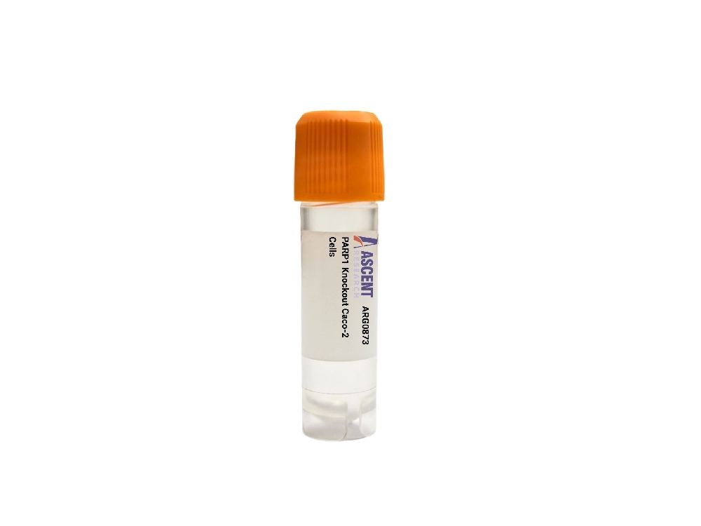
Genome-edited Cells
Large intestine (colon)
The PARP1 Knockout Caco-2 Cells are a CRISPR/Cas9-edited colorectal adenocarcinoma cell line with targeted disruption of the PARP1 gene. Derived from the well-characterized Caco-2 intestinal epithelial model, this loss-of-function line enables precise investigation of DNA repair, cell death, and inflammatory signaling pathways in an absorptive enterocyte-like context. PARP1 is a key sensor of DNA strand breaks and catalyzes PARylation of target proteins including XRCC1, p53, and histones, with hyperactivation leading to NAD+ depletion and parthanatos. This knockout model is ideal for studying DNA damage responses, screening PARP inhibitors, and assessing intestinal toxicity in drug discovery research.
 BMT2 Knockout HAP1 Polyclonal Cells
Cat. No. ARG22109
FOXK2 Knockout A2780 Polyclonal Cells
Cat. No. ARG18865
BCL7A Knockout HGC-27 Polyclonal Cells
Cat. No. ARG29709
EZR Knockout 786-O Polyclonal Cells
Cat. No. ARG5598
BMT2 Knockout HAP1 Polyclonal Cells
Cat. No. ARG22109
FOXK2 Knockout A2780 Polyclonal Cells
Cat. No. ARG18865
BCL7A Knockout HGC-27 Polyclonal Cells
Cat. No. ARG29709
EZR Knockout 786-O Polyclonal Cells
Cat. No. ARG5598
 Pig Prostate Fibroblast Medium
Cat. No. ARM0916
Pig Prostate Fibroblast Medium
Cat. No. ARM0916
 Rabbit Thyroid Epithelial Cell Medium
Cat. No. ARM0757
Rabbit Thyroid Epithelial Cell Medium
Cat. No. ARM0757
The PARP1 Knockout Caco-2 Cells product is a CRISPR/Cas9-edited knockout cell line derived from the Caco-2 human colorectal adenocarcinoma line, engineered to disrupt the PARP1 gene through targeted gene inactivation. This loss-of-function model eliminates wild-type PARP1 expression, enabling precise investigation of PARP1-dependent molecular processes without interference from endogenous poly(ADP-ribose) polymerase 1 activity.
The parental Caco-2 cell line was originally isolated from a 72-year-old Caucasian male with colorectal adenocarcinoma and has become a gold-standard in vitro model for intestinal epithelial research. Upon reaching confluence, Caco-2 cells differentiate spontaneously into a polarized monolayer with morphological and functional characteristics of small intestinal absorptive enterocytes, including the formation of tight junctions, apical brush border microvilli, and expression of key drug-metabolizing enzymes and transporters. This makes them invaluable for studies on intestinal drug absorption, metabolism, and the epithelial barrier function.
PARP1 is a nuclear enzyme that senses DNA single-strand breaks and catalyzes poly(ADP-ribose) (PAR) synthesis from NAD+, modifying itself and target proteins such as histones, p53, XRCC1, and DNA ligase III. Its activity is regulated by DNA damage, reactive oxygen species, NAD+ levels, and upstream signals from sirtuins, p53, and BRCA1. PARP1 interacts directly with XRCC1, DNA ligase III, PARP2, APE1, and PARG to assemble repair complexes at damage sites, while downstream PARylation influences NF-??B transcriptional activity and p53 stability, linking PARP1 to apoptosis, necroptosis, and inflammatory signaling. Excessive PARP1 activation depletes cellular NAD+ and ATP, driving parthanatos via mitochondrial apoptosis-inducing factor (AIF) release.
In the context of Caco-2 cells, PARP1 knockout provides a unique platform to dissect DNA damage responses within the intestinal epithelium. Given the critical barrier function of these cells, PARP1 deficiency allows researchers to explore the role of PARylation in maintaining epithelial integrity under genotoxic or inflammatory stress, as may occur in chemotherapy-induced mucositis, inflammatory bowel disease, or ischemia-reperfusion injury. Additionally, this model can be used to investigate how loss of PARP1 affects the expression and functionality of drug-metabolizing enzymes and transporters, potentially altering intestinal drug absorption profiles.
This knockout cell line is suitable for a broad range of applications, including mechanistic studies of base excision repair and single-strand break repair, evaluation of PARP inhibitor efficacy and resistance mechanisms in colorectal adenocarcinoma, and screening of compounds that modulate parthanatos or necroptosis. Researchers can employ assays such as Western blotting for residual PARP1 and auto-ADP-ribosylation, RT-qPCR for transcript analysis, immunofluorescence for subcellular localization, comet assays for DNA damage, MTT or Alamar blue viability assays, NAD+ quantification, and PARylation activity measurements. For further technical details or to discuss custom requirements, please contact Ascent Research.