Genome-edited Cells
Embryo
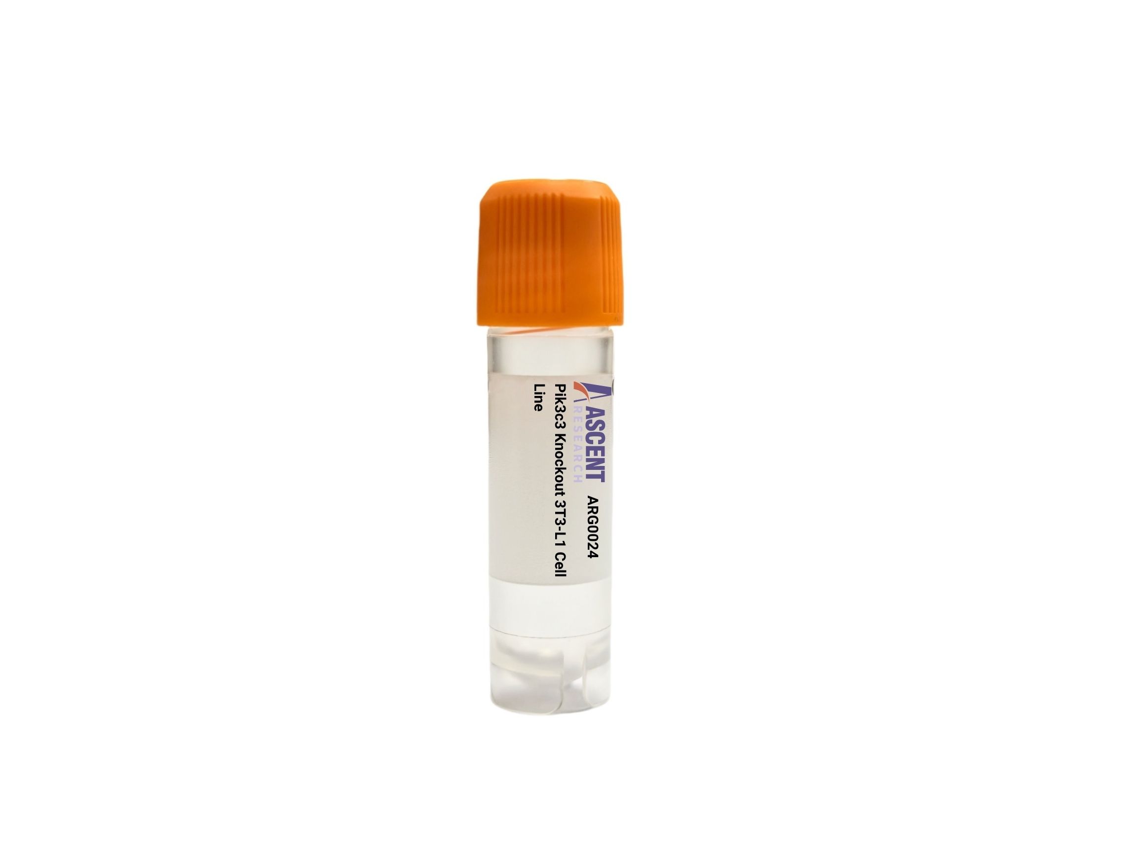
Pik3c3 Knockout 3T3-L1 is a CRISPR/Cas9-edited mouse preadipocyte cell line with disruption of Pik3c3, which encodes VPS34, the class III PI3K that produces phosphatidylinositol 3-phosphate. In 3T3-L1 cells, this model supports analysis of autophagy initiation, endosomal trafficking, lysosomal homeostasis, adipocyte differentiation, and metabolic stress responses. VPS34 functions in complexes with BECN1, ATG14, UVRAG, and PIK3R4/VPS15 and acts upstream of WIPI2 recruitment, LC3 lipidation, and SQSTM1 turnover. Applications include autophagic flux assays, LC3 and LAMP1 imaging, Oil Red O staining, insulin/metabolic studies, and phosphoinositide signaling research.
 CALD1 Knockout A2780 Polyclonal Cells
Cat. No. ARG28981
MKI67 Knockout A2780 Polyclonal Cells
Cat. No. ARG18793
ANO6 Knockout HT29 Polyclonal Cells
Cat. No. ARG32955
CD274 Knockout HT29 Polyclonal Cells
Cat. No. ARG43436
LCN2 Knockout MCF7 Polyclonal Cells
Cat. No. ARG11422
NTMT1 Knockout MES-OV Polyclonal Cells
Cat. No. ARG6219
CALD1 Knockout A2780 Polyclonal Cells
Cat. No. ARG28981
MKI67 Knockout A2780 Polyclonal Cells
Cat. No. ARG18793
ANO6 Knockout HT29 Polyclonal Cells
Cat. No. ARG32955
CD274 Knockout HT29 Polyclonal Cells
Cat. No. ARG43436
LCN2 Knockout MCF7 Polyclonal Cells
Cat. No. ARG11422
NTMT1 Knockout MES-OV Polyclonal Cells
Cat. No. ARG6219
The Pik3c3 Knockout 3T3-L1 Cell Line is a CRISPR/Cas9-engineered mouse cell model in which the Pik3c3 gene has been disrupted to abolish functional expression of the class III phosphatidylinositol 3-kinase catalytic subunit VPS34. This stable gene-edited in vitro system is generated in 3T3-L1 cells, a fibroblast-like preadipocyte cell line, and is designed for mechanistic studies of autophagy, endomembrane trafficking, lysosomal biology, and metabolic regulation in a well-established adipocyte precursor background.
3T3-L1 cells are derived from murine embryonic fibroblast-like precursors and are widely used because they undergo hormonally induced differentiation into adipocyte-like cells with robust lipid accumulation and insulin-responsive glucose metabolism. As a result, this host line is a standard experimental platform for investigating adipogenesis, lipid droplet formation, adipokine-related biology, nutrient sensing, and cellular responses relevant to obesity, insulin resistance, type 2 diabetes, and fatty liver disease. The model also provides a tractable system for analyzing autophagy-dependent remodeling events that accompany differentiation and metabolic adaptation.
PIK3C3/VPS34 is the catalytic core of class III PI3K complexes that generate phosphatidylinositol 3-phosphate on nascent autophagic and endosomal membranes. Its activity is regulated by nutrient deprivation, amino acid availability, AMPK, MTOR, and ULK1, and it functions in multiprotein complexes with PIK3R4/VPS15, BECN1, ATG14, UVRAG, RUBICON, NRBF2, and AMBRA1. Through these assemblies, VPS34 acts upstream of WIPI2 recruitment, DFCP1-positive omegasome formation, LC3 lipidation, SQSTM1/p62 turnover, autophagosome formation, endosome maturation, and lysosome-dependent cargo degradation. Because these processes are central to membrane dynamics and stress adaptation, Pik3c3 loss is relevant to metabolic disease, neurodegeneration, cancer, and lysosomal dysfunction research.
In the 3T3-L1 context, Pik3c3 disruption provides a useful model to examine how defective phosphatidylinositol 3-phosphate production alters preadipocyte homeostasis and adipocyte differentiation-associated remodeling. The model can support studies on how autophagosome nucleation and endolysosomal trafficking influence lipid storage, insulin-responsive pathways, and nutrient stress responses in adipocyte-lineage cells. It is also suitable for probing pathway dependence within BECN1-ATG14 or UVRAG-containing VPS34 complexes and for comparing autophagy-linked versus endosomal functions during metabolic adaptation.
Representative applications include western blot analysis of LC3-I/LC3-II conversion and SQSTM1 accumulation, immunofluorescence imaging of LC3, WIPI2, and LAMP1 localization, autophagic flux studies using lysosomal inhibitors, and phosphatidylinositol 3-phosphate reporter imaging. Researchers may also combine this model with Oil Red O staining, RT-qPCR of adipogenic and stress-response genes, RNA-seq, glucose uptake or mitochondrial function assays, electron microscopy of autophagic structures, and co-immunoprecipitation of VPS34 complex components to define Pik3c3-dependent signaling and trafficking mechanisms. Researchers may contact Ascent Research for additional technical information, product details, or related gene-edited cell models.