Genome-edited Cells
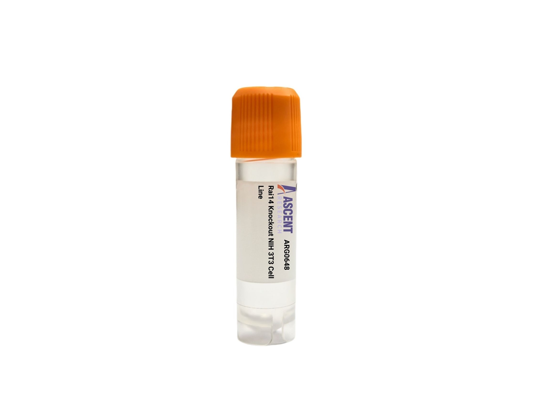
The Rai14 Knockout NIH 3T3 Cell Line is a CRISPR/Cas9-edited mouse embryonic fibroblast line with targeted disruption of the Rai14 gene, encoding the actin scaffold protein RAI14. This loss-of-function model is derived from the immortalized NIH 3T3 host, a standard system for studying cytoskeletal organization and cell motility. RAI14 crosslinks actin filaments and governs focal adhesion dynamics via interactions with FAK, ??-catenin, and Rho GTPase?CROCK pathways. The knockout line is designed for applications in cancer metastasis, spermatogenesis, and adhesion research, enabling assays such as wound healing, Transwell migration, and immunodetection of actin and phospho-FAK.
 ICMT Knockout K562 Polyclonal Cells
Cat. No. ARG20621
Map4k1 Knockout RAW 264.7 Polyclonal Cells
Cat. No. ARG12441
GRHL1 Knockout HAP1 Polyclonal Cells
Cat. No. ARG22378
BLOC1S2 Knockout 786-O Polyclonal Cells
Cat. No. ARG25088
ACTR1B Knockout HEK293T Polyclonal Cells
Cat. No. ARG38206
ICMT Knockout K562 Polyclonal Cells
Cat. No. ARG20621
Map4k1 Knockout RAW 264.7 Polyclonal Cells
Cat. No. ARG12441
GRHL1 Knockout HAP1 Polyclonal Cells
Cat. No. ARG22378
BLOC1S2 Knockout 786-O Polyclonal Cells
Cat. No. ARG25088
ACTR1B Knockout HEK293T Polyclonal Cells
Cat. No. ARG38206
 J82
Cat. No. ARC0362
J82
Cat. No. ARC0362
The Rai14 Knockout NIH 3T3 Cell Line is a CRISPR/Cas9-edited knockout cell line derived from the spontaneously immortalized mouse embryonic fibroblast line NIH 3T3. This model was generated via CRISPR/Cas9-mediated disruption of the Rai14 gene, which encodes the RAI14 scaffold protein. The resulting cell line serves as a loss-of-function tool to study RAI14-dependent processes in a controlled in vitro setting.
NIH 3T3 cells are a widely used fibroblast line established from disaggregated Swiss mouse embryos, known for their robust growth, contact inhibition, and suitability for studies of cytoskeletal dynamics, adhesion, and motility. As spontaneously immortalized cells, they provide a stable and easily transfectable host for gene-editing applications, enabling consistent phenotypic analyses.
RAI14, also known as retinoic acid-induced protein 14, is an ankyrin repeat-containing scaffold protein that crosslinks actin filaments and coordinates focal adhesion (FA) turnover. It interacts with actin, filamin, cortactin, and ARPC2 to stabilize the cortical actin network. Upstream, RAI14 is regulated by retinoic acid signaling and the Wnt pathway, while downstream it modulates actin filament organization, FAK phosphorylation, and ??-catenin activity. RAI14 integrates signals from Rho GTPases, ROCK, FAK, Src, and paxillin to control actin remodeling, cell adhesion, and migration.
In the NIH 3T3 background, disruption of Rai14 impairs actin cytoskeleton organization, leading to reduced migration velocity and altered focal adhesion dynamics. This phenotype recapitulates key features of RAI14 loss observed in other cell types and provides a physiologically relevant fibroblast model for dissecting the molecular mechanisms underlying cell motility and adhesion. Because NIH 3T3 cells are of mesenchymal origin, the knockout line is particularly valuable for investigating pathways that drive fibroblast migration during wound healing and cancer metastasis.
This knockout cell line is ideally suited for a variety of functional assays, including Western blotting to assess FAK phosphorylation and actin levels, immunofluorescence staining for F-actin to visualize cytoskeletal changes, wound healing and Transwell migration assays to quantify motility deficits, co-immunoprecipitation to map RAI14-interacting protein complexes, and RT-qPCR to evaluate expression of downstream targets such as ??-catenin. Researchers can employ this model to study actin cytoskeleton remodeling, focal adhesion signaling, and the molecular basis of cancer cell dissemination or spermatogenesis defects. For further information, please contact Ascent Research.