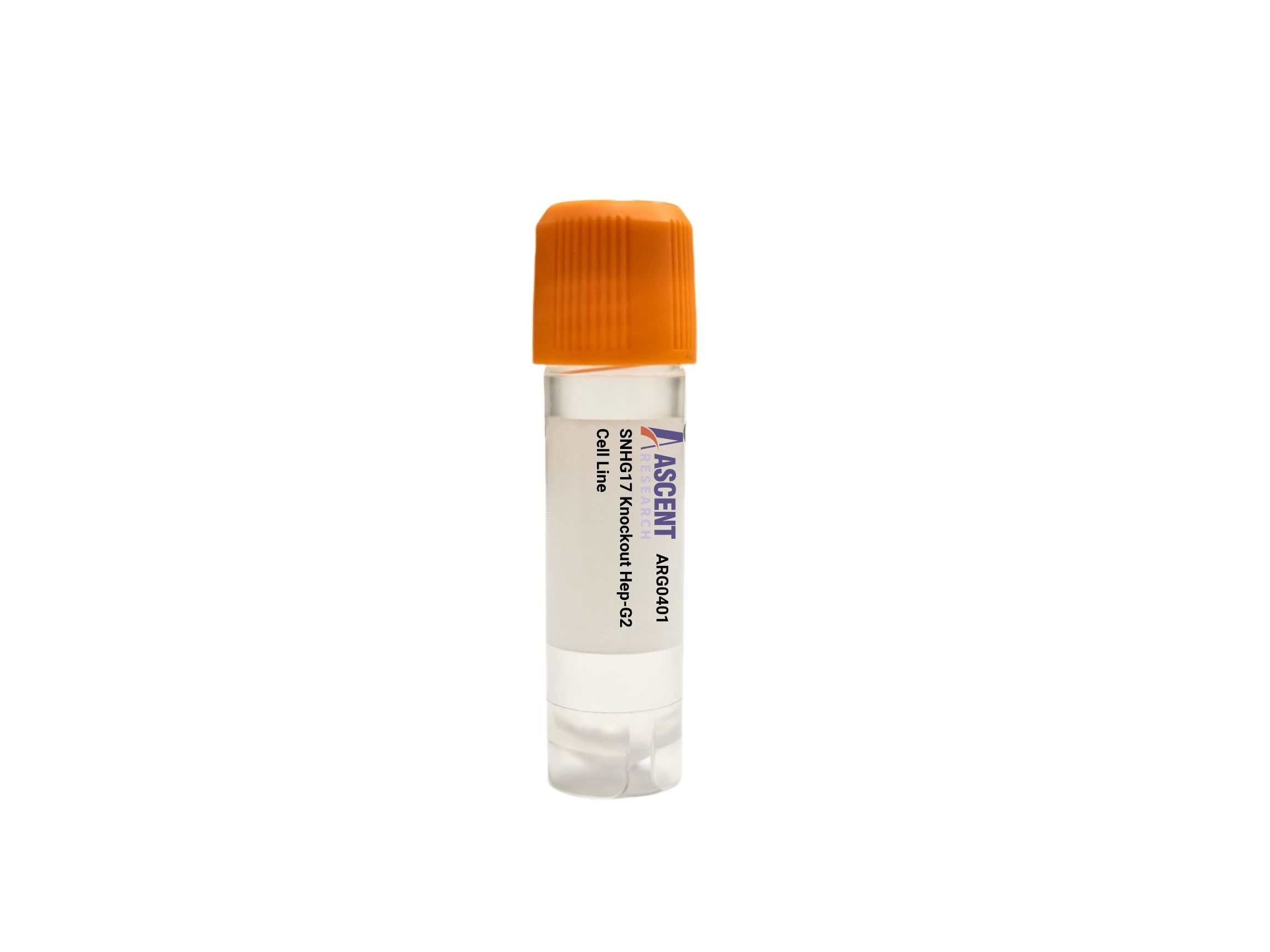
Genome-edited Cells
Liver
The SNHG17 Knockout Hep-G2 Cell Line offers a CRISPR/Cas9-edited loss-of-function model in human hepatocellular carcinoma cells. This product enables targeted disruption of the oncogenic lncRNA SNHG17, which promotes liver cancer through ceRNA-mediated regulation of miRNAs and downstream targets. SNHG17, activated by SP1, STAT3, and c-MYC, sponges miR-338-3p, miR-186-5p, and miR-485-5p to upregulate SOX4, ZEB1, and Cyclin D1, driving PI3K/AKT and Wnt/??-catenin signaling. The knockout cell line is ideal for proliferation, migration, invasion, and apoptosis assays, supporting research in ceRNA networks, drug target validation, and biomarker discovery.
 LARP1B Knockout A2780 Polyclonal Cells
Cat. No. ARG18575
LARP1B Knockout A2780 Polyclonal Cells
Cat. No. ARG18575
 RNF165 Knockout Huh-7 Cell Line
Cat. No. ARG44091
GPX1 Knockout HEK293T Polyclonal Cells
Cat. No. ARG26011
JAG2 Knockout KYSE150 Polyclonal Cells
Cat. No. ARG36257
LCLAT1 Knockout Hela Polyclonal Cells
Cat. No. ARG7853
FKBP5 Knockout AGS Polyclonal Cells
Cat. No. ARG3062
RNF165 Knockout Huh-7 Cell Line
Cat. No. ARG44091
GPX1 Knockout HEK293T Polyclonal Cells
Cat. No. ARG26011
JAG2 Knockout KYSE150 Polyclonal Cells
Cat. No. ARG36257
LCLAT1 Knockout Hela Polyclonal Cells
Cat. No. ARG7853
FKBP5 Knockout AGS Polyclonal Cells
Cat. No. ARG3062
The SNHG17 Knockout Hep-G2 Cell Line is a CRISPR/Cas9-edited knockout cell line derived from Homo sapiens hepatocellular carcinoma Hep-G2 cells. This loss-of-function model enables targeted disruption of the SNHG17 long non-coding RNA gene, providing a reliable system for investigating its roles in cancer. The cell line is maintained under standard culture conditions, ready for functional genomics and signaling studies.
The Hep-G2 host cell line originates from a liver biopsy of a 15-year-old male with hepatocellular carcinoma. These epithelial cells retain key hepatic functions, including drug metabolism and hepatotoxicity responses, making them a widely used model for liver biology. Hep-G2 cells express relevant cytochrome P450 enzymes and respond to genotoxic and metabolic stresses, offering a physiologically relevant context for studying hepatic carcinogenesis and compound safety.
SNHG17 functions as an oncogenic lncRNA that drives tumor cell proliferation, migration, and invasion primarily through a ceRNA mechanism. Upstream transcription factors SP1, STAT3, and c-MYC activate its expression. SNHG17 sponges tumor-suppressive miRNAs such as miR-338-3p, miR-186-5p, and miR-485-5p, thereby derepressing targets like SOX4, ZEB1, Cyclin D1, and Bcl-2. This network converges on PI3K/AKT and Wnt/??-catenin signaling, with representative components PIK3CA, AKT1, mTOR, CTNNB1, and TCF4, as well as EMT regulators CDH1, VIM, and SNAI1.
In Hep-G2 cells, SNHG17 knockout attenuates oncogenic signaling and allows dissection of lncRNA-mediated tumor progression. Loss of SNHG17 is anticipated to reduce PI3K/AKT and Wnt/??-catenin pathway activity, impair migration and invasion, and alter apoptosis and cell cycle profiles. This model facilitates validation of miRNA-lncRNA interactions and EMT regulation, and it can be used to evaluate whether Hep-G2 tumorigenicity depends on SNHG17.
The cell line supports multiple functional assays: MTT/CCK-8 proliferation, colony formation, wound healing, Transwell migration/invasion, flow cytometry for apoptosis and cell cycle, RT-qPCR, and Western blot for targets like Cyclin D1 and ZEB1. Dual luciferase reporter assays confirm miRNA interactions. Applications include lncRNA functional studies, ceRNA network investigation, drug target validation, and biomarker discovery in liver cancer. For further inquiries, contact Ascent Research.