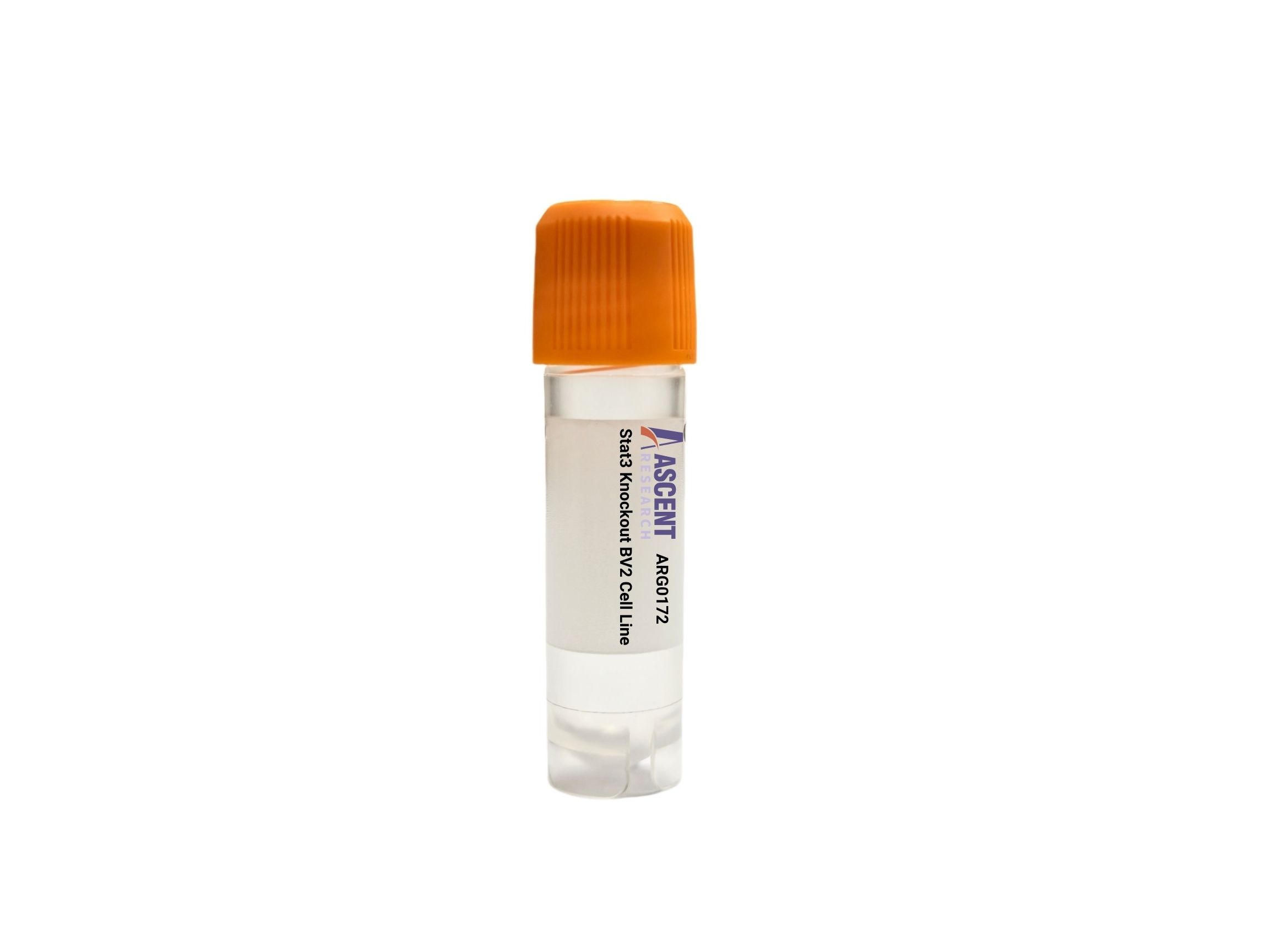
Genome-edited Cells
Stat3 Knockout BV2 is a CRISPR/Cas9-engineered mouse microglia-like cell line with disruption of the Stat3 gene, providing a stable in vitro model for neuroinflammation and innate immune signaling studies. In BV2 cells, STAT3 functions downstream of IL6R/gp130 and JAK1/JAK2/TYK2 and intersects with TLR4/NFKB crosstalk to regulate targets such as Socs3, Ccl2, Il6, and Il10. This model is useful for microglial activation analysis, cytokine signaling studies, transcriptional target validation, and drug response testing using western blotting, RT-qPCR, RNA-seq, ELISA, phospho-STAT3 assays, flow cytometry, and phagocytosis assays.
 GPAM Knockout HAP1 Polyclonal Cells
Cat. No. ARG22293
LMNB1 Knockout 143B Polyclonal Cells
Cat. No. ARG11887
GPR108 Knockout MES-OV Polyclonal Cells
Cat. No. ARG24465
EFHD1 Knockout NCI-H1299 Polyclonal Cells
Cat. No. ARG40655
EEF1A2 Knockout NCI-H1299 Polyclonal Cells
Cat. No. ARG40549
GPAM Knockout HAP1 Polyclonal Cells
Cat. No. ARG22293
LMNB1 Knockout 143B Polyclonal Cells
Cat. No. ARG11887
GPR108 Knockout MES-OV Polyclonal Cells
Cat. No. ARG24465
EFHD1 Knockout NCI-H1299 Polyclonal Cells
Cat. No. ARG40655
EEF1A2 Knockout NCI-H1299 Polyclonal Cells
Cat. No. ARG40549
 Rabbit Renal Interstitial Fibroblasts
Cat. No. ARP0674
Rabbit Renal Interstitial Fibroblasts
Cat. No. ARP0674
The Stat3 Knockout BV2 Cell Line is a CRISPR/Cas9-engineered murine microglia-like cell model in which the Stat3 gene has been disrupted to eliminate functional STAT3 expression. This stable knockout line provides an in vitro system for interrogating STAT3-dependent signaling and transcriptional regulation in cells with key features of central nervous system innate immune effectors. Because BV2 cells are widely used to model activated microglia, this gene-edited derivative is suited for mechanistic studies of inflammatory signaling, cytokine responsiveness, and microglial state regulation.
BV2 is an immortalized mouse microglia-like cell line broadly used as a tractable experimental model of CNS resident macrophage-like cells. It is relevant to studies of neuroinflammation, phagocytosis, cytokine production, and neuron-glia interactions, and has been extensively applied in research on Alzheimer??s disease, Parkinson??s disease, multiple sclerosis, ischemic brain injury, CNS infection, neuropathic pain, and glioma-associated immune signaling. In culture, BV2 cells support controlled analysis of stimulus-dependent innate immune programs, including responses to inflammatory cytokines and TLR4 activation, making them useful for dissecting regulatory pathways that influence microglial activation state.
STAT3 is a latent cytoplasmic transcription factor that functions downstream of multiple cytokine and growth factor receptors. It is activated primarily by tyrosine phosphorylation mediated by JAK1, JAK2, and TYK2 following signaling through IL6R and gp130/IL6ST, and can also be engaged downstream of IFNAR, EGFR, CSF1R, Src family kinases, and TLR4 stimulation by LPS through pathway crosstalk. Upon phosphorylation, STAT3 dimerizes, translocates to the nucleus through importin alpha/beta-dependent transport, and transcriptionally regulates targets including Socs3, Bcl2l1, Myc, Ccnd1, Vegfa, Ccl2, Il6, and Il10. STAT3 signaling intersects functionally with STAT1, NFKB p65/RelA, ERK1/2, AKT, CBP/EP300, PIAS3, and SOCS3, placing it at a central node linking JAK-STAT signaling to NF-kB-, MAPK-, and neuroinflammation-associated transcriptional programs.
In the BV2 background, loss of STAT3 is a useful model for evaluating how cytokine-responsive transcription contributes to microglial inflammatory output, survival pathways, and activation-state transitions. This system can support studies of canonical IL-6 family signaling as well as crosstalk between JAK-STAT3 and TLR4/NFKB pathways, with relevance to disease settings in which microglial activation shapes tissue damage, repair, or tumor-associated immune phenotypes.
The Stat3 knockout BV2 model can be applied to phospho-signaling studies by western blotting, transcriptional profiling by RT-qPCR or RNA-seq, and cytokine output analysis by ELISA following stimulation with IL6, LPS, interferon-related ligands, or growth factor inputs. It is also suitable for immunofluorescence-based localization studies, reporter assays, ChIP-qPCR for STAT3-regulated loci, co-immunoprecipitation of interacting signaling components, flow cytometric phenotyping, phagocytosis assays, apoptosis assays, and cell viability measurements to define pathway dependency and inflammatory response programs. Researchers may contact Ascent Research for additional technical information, product details, or related gene-edited cell models.