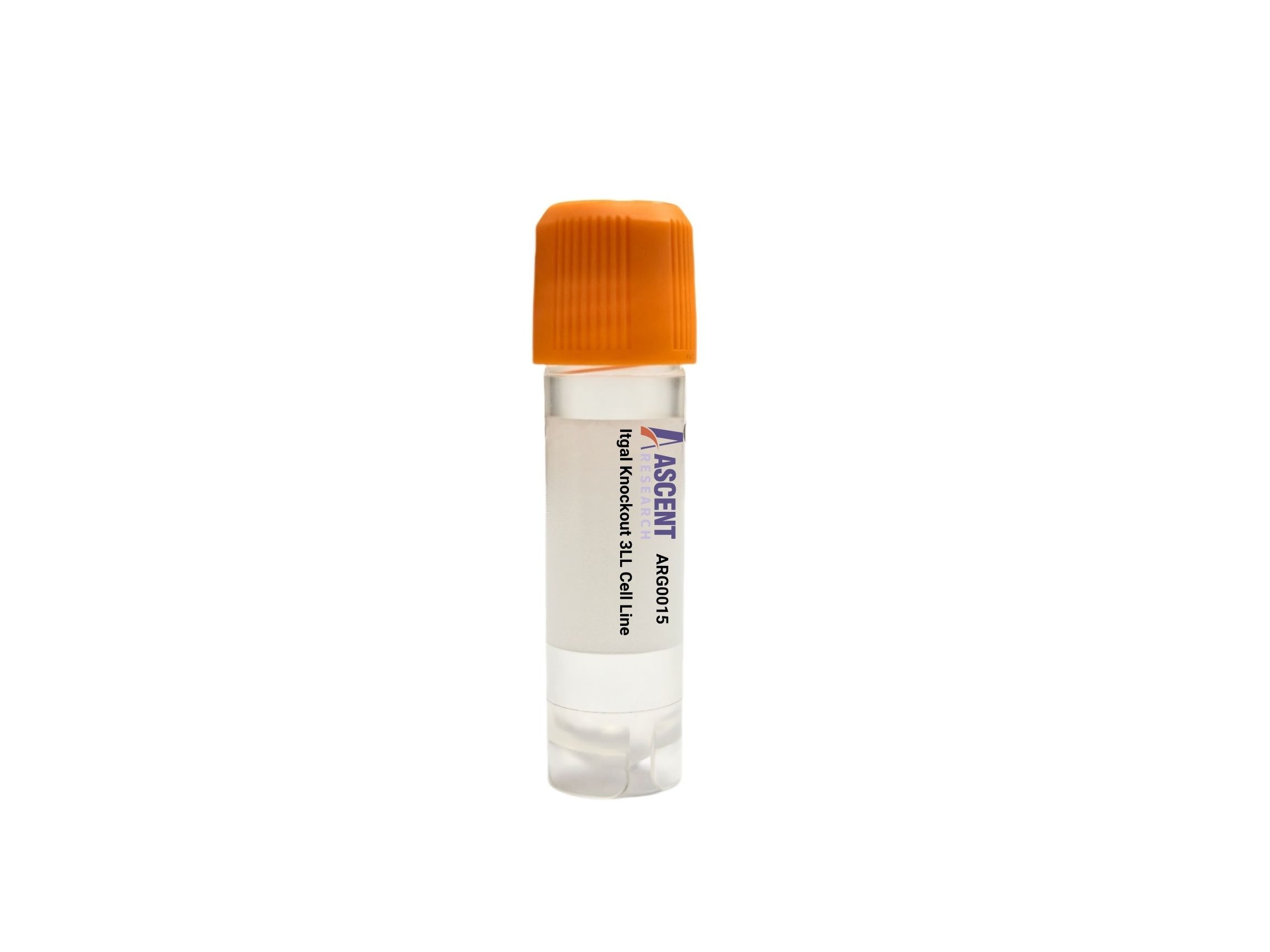
Genome-edited Cells
Lung
The Itgal Knockout 3LL Cell Line is a CRISPR/Cas9-edited mouse Lewis lung carcinoma model with disruption of Itgal, which encodes integrin alpha L (CD11a), the alpha subunit of LFA-1. In 3LL cells, loss of ITGAL enables analysis of ICAM1/ICAM2-dependent adhesion, ITGB2-associated integrin function, and downstream signaling involving FAK, SRC, RAC1, and actin remodeling. This syngeneic C57BL/6-compatible tumor model is useful for tumor-immune interaction studies, adhesion and migration assays, co-culture experiments with leukocytes, phospho-signaling analysis, and metastasis or immune evasion research in immuno-oncology settings.
 CASP3 Knockout A549 Polyclonal Cells
Cat. No. ARG32009
MYH10 Knockout A2780 Polyclonal Cells
Cat. No. ARG18651
FIBP Knockout NCI-H1975 Polyclonal Cells
Cat. No. ARG16513
CNP Knockout NCI-H1299 Polyclonal Cells
Cat. No. ARG17585
HEBP2 Knockout HT29 Polyclonal Cells
Cat. No. ARG33332
CASP3 Knockout A549 Polyclonal Cells
Cat. No. ARG32009
MYH10 Knockout A2780 Polyclonal Cells
Cat. No. ARG18651
FIBP Knockout NCI-H1975 Polyclonal Cells
Cat. No. ARG16513
CNP Knockout NCI-H1299 Polyclonal Cells
Cat. No. ARG17585
HEBP2 Knockout HT29 Polyclonal Cells
Cat. No. ARG33332
 Mouse Hippocampal Neuron Medium
Cat. No. ARM0610
Mouse Hippocampal Neuron Medium
Cat. No. ARM0610
The Itgal Knockout 3LL Cell Line is a CRISPR/Cas9-engineered mouse Lewis lung carcinoma model in which the Itgal gene has been disrupted to eliminate functional integrin alpha L expression. This gene-edited cell line provides a stable in vitro system for examining the consequences of Itgal loss in a tumor epithelial-like background. As ITGAL encodes CD11a, the alpha subunit of lymphocyte function-associated antigen-1 (LFA-1), this model is suited for studies of adhesion-dependent signaling, cell-cell interactions, and tumor-associated mechanisms influenced by integrin-mediated communication.
3LL, also known as Lewis lung carcinoma, is a murine lung carcinoma cell line syngeneic to C57BL/6 mice and is extensively used in studies of lung cancer progression, metastatic dissemination, and tumor-immune interactions. Its broad use in immuno-oncology derives from its compatibility with syngeneic mouse tumor models and its utility in evaluating tumor cell behavior within immune-relevant experimental settings. In vitro, 3LL cells serve as a practical platform for investigating migration, invasion, and responses to stromal or immune cues, making them a useful host for dissecting adhesion and signaling pathways linked to cancer biology.
At the molecular level, ITGAL forms a heterodimer with ITGB2 to generate LFA-1, a receptor that mediates ICAM-dependent adhesion through binding to ICAM1 and ICAM2. ITGAL function is regulated by inside-out activation pathways downstream of chemokine receptors, including CXCR4 and CCR7, and by RAP1A, APBB1IP/RIAM, TLN1, and FERMT3, which promote integrin activation and ligand binding competence. Following ligand engagement, LFA-1 mediates outside-in signaling involving PTK2/FAK, SRC family kinases, PI3K-AKT, ERK, RAC1 activation, and actin remodeling through cytoskeletal components including ACTB and filamin A, with paxillin-associated adhesive signaling contributing to migratory phenotypes. Disruption of Itgal is therefore expected to diminish LFA-1-dependent adhesive interactions and alter downstream signaling relevant to immune synapse organization, transendothelial migration biology, and cytoskeletal regulation.
In the 3LL background, loss of Itgal provides a means to interrogate how integrin alpha L contributes to tumor cell interaction with immune and microenvironmental ligands, as well as how reduced ICAM-directed adhesion influences migration-associated and signaling-associated phenotypes. This context is relevant to studies of tumor immune evasion, inflammatory signaling interfaces, and metastatic behavior, particularly where adhesion receptor networks affect contact-dependent communication with leukocytes.
This knockout model can be applied in flow cytometry assays evaluating CD11a/LFA-1 surface expression, RT-qPCR or western blotting to confirm Itgal disruption, and adhesion assays using ICAM-coated substrates to quantify changes in ligand-dependent attachment. It is also suitable for migration and invasion assays, immunofluorescence analysis of actin cytoskeletal organization, phospho-signaling studies focused on FAK, SRC, AKT, or ERK pathway activity, and co-culture experiments with T cells or NK cells to examine tumor-immune engagement. In syngeneic mouse studies and transcriptomic profiling by RNA-seq, the model may support investigation of adhesion-regulated programs and integrin pathway dependencies in cancer immunology and metastasis research. Researchers may contact Ascent Research for additional technical information, product details, or related gene-edited cell models.