Genome-edited Cells
Breast (mammary gland)
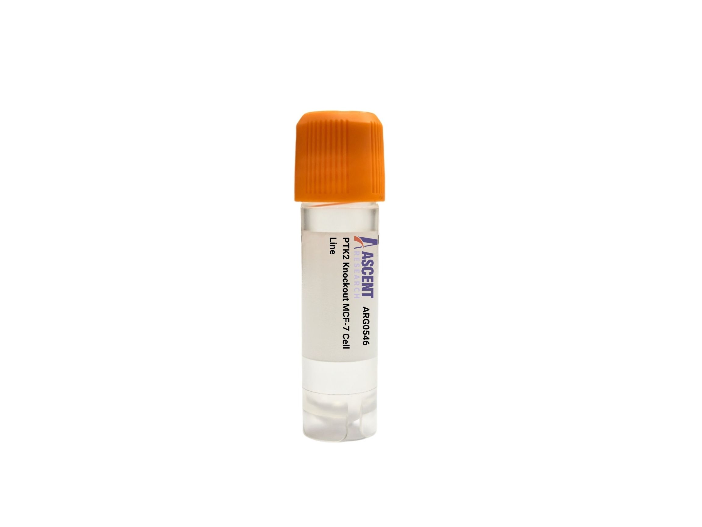
The PTK2 Knockout MCF-7 Cell Line is a CRISPR/Cas9-edited human breast cancer cell model with targeted disruption of the PTK2 gene, eliminating expression of focal adhesion kinase (FAK). Derived from the ER+, PR+, HER2? MCF-7 breast adenocarcinoma cell line, this loss-of-function model enables precise dissection of FAK-dependent signaling in a hormone-responsive background. FAK is a key mediator of integrin and growth factor receptor signaling, regulating cell adhesion, migration, survival, and proliferation through downstream effectors such as Src, PI3K/AKT, and MAPK/ERK. This knockout cell line is ideal for investigating breast cancer metastasis, drug resistance, and adhesion dynamics using standard assays including western blotting, transwell migration, colony formation, and immunofluorescence.
 KIAA0930 Knockout SiHa Cell Line
Cat. No. ARG43937
KIAA0930 Knockout SiHa Cell Line
Cat. No. ARG43937
 AP4B1 Knockout 786-O Polyclonal Cells
Cat. No. ARG24932
HEXB Knockout NCI-H1975 Polyclonal Cells
Cat. No. ARG31623
FAM131C Knockout HEK293T Polyclonal Cells
Cat. No. ARG3432
MS4A1 Knockout Raji Polyclonal Cells
Cat. No. ARG1275
AP4B1 Knockout 786-O Polyclonal Cells
Cat. No. ARG24932
HEXB Knockout NCI-H1975 Polyclonal Cells
Cat. No. ARG31623
FAM131C Knockout HEK293T Polyclonal Cells
Cat. No. ARG3432
MS4A1 Knockout Raji Polyclonal Cells
Cat. No. ARG1275
 Mouse Vertebral Endplate Chondrocytes
Cat. No. ARP0572
Mouse Vertebral Endplate Chondrocytes
Cat. No. ARP0572
The PTK2 Knockout MCF-7 Cell Line is a genetically engineered human cell model featuring CRISPR/Cas9-mediated disruption of the PTK2 gene, which encodes focal adhesion kinase (FAK). This knockout cell line provides researchers with a targeted loss-of-function system to dissect FAK-dependent signaling networks in a well-characterized epithelial breast cancer background. The gene disruption is introduced in the MCF-7 host cell line, enabling comparative studies with wild-type controls to elucidate FAK??s contributions to adhesion, migration, survival, and proliferation. The product is supplied as a live cell line and is suitable for immediate expansion in standard culture conditions.
The MCF-7 host cell line is derived from human breast adenocarcinoma and represents an estrogen receptor-positive (ER+), progesterone receptor-positive (PR+), and HER2-negative (HER2?) phenotype. This cell line is a foundational model for hormone-responsive breast cancer research, widely employed to investigate endocrine therapy resistance, metastatic progression, and tumor cell biology. MCF-7 cells retain epithelial characteristics and form polarized monolayers, making them particularly valuable for studying cell adhesion, junctional complexes, and cytoskeletal dynamics. Their well-documented signaling landscape and genetic tractability further enhance the utility of knockout derivatives for mechanistic and pharmacological studies.
PTK2 encodes FAK, a non-receptor tyrosine kinase that functions as a central mediator of integrin and growth factor receptor signaling. FAK is activated in response to integrin clustering, extracellular matrix (ECM) engagement by fibronectin, and stimulation of receptor tyrosine kinases such as EGFR and HER2, as well as G-protein coupled receptors and the pro-inflammatory cytokine TNF??. Mechanical stress also serves as an upstream input. Upon activation, FAK autophosphorylates at Y397, creating a high-affinity binding site for Src family kinases. The resultant FAK?CSrc complex orchestrates a signaling platform involving adaptor proteins p130Cas and paxillin, and guanine nucleotide exchange factors such as Sos. This complex propagates signals through the PI3K/AKT and MAPK/ERK cascades, leading to downstream effects on cell cycle regulators (Cyclin D1) and apoptosis regulators (Bcl-2). Additionally, FAK modulates Rho family GTPases including Rac and Rho, thereby influencing actin cytoskeleton reorganization and focal adhesion dynamics.
In the MCF-7 breast cancer cell context, FAK signaling is intricately linked to estrogen-independent growth, survival, and metastatic dissemination. Disruption of PTK2 abrogates key integrin-mediated adhesive and migratory responses, and attenuates signaling through the PI3K/AKT and MAPK/ERK pathways that are frequently co-opted in endocrine-resistant disease. Consequently, this knockout model enables detailed examination of FAK??s role in promoting anchorage-independent growth, a hallmark of metastatic competence, and in mediating crosstalk with estrogen receptor signaling. As a result, the PTK2 Knockout MCF-7 Cell Line is a powerful tool for investigating mechanisms of tumor progression, resistance to anti-estrogen therapies, and tumor angiogenesis, where FAK activity is often dysregulated.
This knockout cell line is ideally suited for a wide array of experimental applications. Researchers can employ western blotting to verify loss of FAK expression and absence of phospho-FAK (Y397) signal, and combine this with phospho-specific antibodies to assess pathway activity. Functional assays include cell adhesion assays on ECM substrates, transwell migration and invasion assays, and colony formation assays to evaluate clonogenic survival. Apoptotic responses can be quantified by Annexin V staining, and immunofluorescence microscopy allows visualization of focal adhesion disassembly and actin stress fiber reorganization. The model is also valuable for drug screening campaigns targeting FAK-dependent vulnerabilities, and for co-culture studies examining tumor?Cstroma interactions. For detailed technical specifications, validation data, or ordering information, please contact Ascent Research.