Genome-edited Cells
Uterus (cervix)
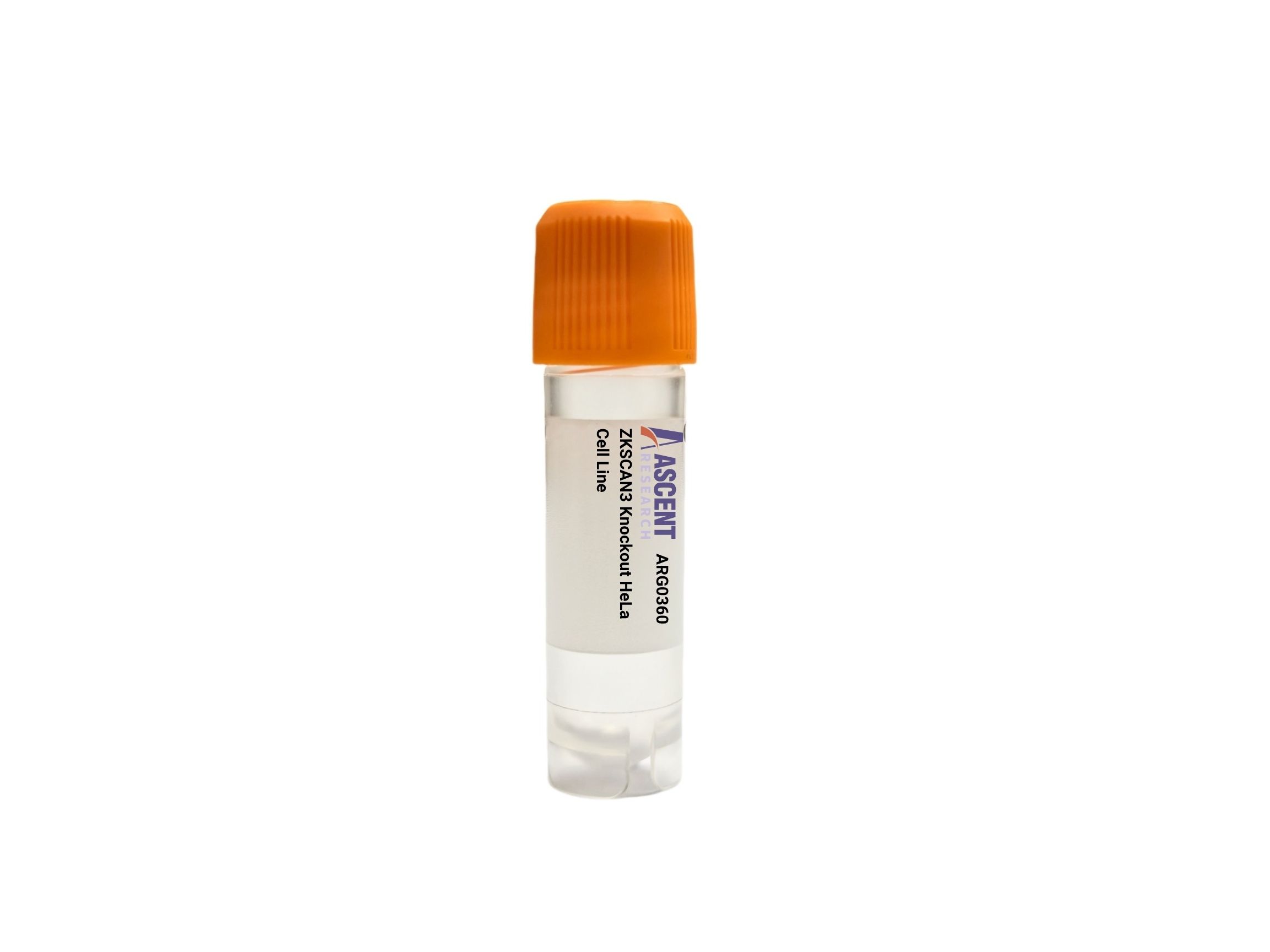
ZKSCAN3 Knockout HeLa Cell Line provides a CRISPR/Cas9-edited loss-of-function model in cervical adenocarcinoma for studying autophagy and lysosomal biogenesis. By disrupting the transcriptional repressor ZKSCAN3, this line derepresses key targets such as MAP1LC3B and LAMP1, enabling investigation of enhanced autophagic flux. Applications include LC3 and p62 western blotting, RT-qPCR, and immunofluorescence for autophagy assessment, as well as lysosomal assays and cell viability tests under stress. The model supports cancer progression, drug screening, and lysosomal disorder research.
 ARRDC1 Knockout HAP1 Polyclonal Cells
Cat. No. ARG21906
ETV6 Knockout A2780 Polyclonal Cells
Cat. No. ARG18567
ETHE1 Knockout jurkat Polyclonal Cells
Cat. No. ARG13573
FILIP1L Knockout A549 Polyclonal Cells
Cat. No. ARG10210
IL12RB2 Knockout HAP1 Polyclonal Cells
Cat. No. ARG22746
CALR Knockout DLD-1 Polyclonal Cells
Cat. No. ARG41890
ARRDC1 Knockout HAP1 Polyclonal Cells
Cat. No. ARG21906
ETV6 Knockout A2780 Polyclonal Cells
Cat. No. ARG18567
ETHE1 Knockout jurkat Polyclonal Cells
Cat. No. ARG13573
FILIP1L Knockout A549 Polyclonal Cells
Cat. No. ARG10210
IL12RB2 Knockout HAP1 Polyclonal Cells
Cat. No. ARG22746
CALR Knockout DLD-1 Polyclonal Cells
Cat. No. ARG41890
The ZKSCAN3 Knockout HeLa Cell Line is a CRISPR/Cas9-edited human cancer model with targeted disruption of the ZKSCAN3 gene. This knockout system eliminates the transcriptional repressor function of ZKSCAN3, enabling investigation of autophagy and lysosomal biogenesis deregulation. Designed for loss-of-function studies, the line permits analysis of pathways normally suppressed by ZKSCAN3 in a widely used cervical adenocarcinoma background.
The parental HeLa cell line is an epithelial cervical carcinoma isolate with a rapid growth rate, adherent morphology, and high transfection efficiency. Its extensive characterization and endogenous expression of essential autophagy and mTOR signaling components provide a reliable platform to examine the consequences of ZKSCAN3 ablation. HeLa cells’ transformed nature also makes this model particularly pertinent for exploring the interplay between oncogenic signaling and autophagic flux.
ZKSCAN3 acts as a master repressor of lysosomal and autophagy-related gene transcription by binding to promoter elements of targets such as MAP1LC3B and LAMP1. mTORC1-dependent phosphorylation regulates its nuclear localization, with active mTORC1 in nutrient-rich conditions maintaining ZKSCAN3’s repressive state. Concurrently, TFEB, a key activator of the same gene set, is reciprocally controlled. SCAND1 and corepressor complexes mediate ZKSCAN3’s silencing functions. Disruption of ZKSCAN3 thus unleashes autophagy and lysosomal gene expression, leading to enhanced degradative capacity independent of TFEB activity.
In HeLa cells, ZKSCAN3 knockout creates a constitutive autophagy-upregulated environment, which can be used to study cancer cell stress adaptation. Enhanced autophagic flux may sensitize or protect cells from chemotherapy, depending on context, and offers a platform for testing compounds that modulate autophagy for therapeutic purposes. The model also serves as a tool to investigate mechanisms underlying lysosomal storage disorders and neurodegeneration where autophagy restoration is beneficial, as it provides a baseline of elevated clearance activity.
Applications include quantitative immunoblotting for LC3-II and p62 to measure autophagic flux, RT-qPCR profiling of autophagy-related transcripts, immunofluorescence for LC3 puncta formation, lysosomal activity assays using fluorogenic substrates, and viability testing under metabolic stress. These assays support cancer research, drug screening for autophagy modulators, and studies of lysosomal dysfunction. For technical details, validation data, or ordering, contact Ascent Research.